Original Article

Isolation and Identification of Fungi Associated with Decay of Quercus mongolica

Youngseok HAM2, Ji-Eun AN2, Soo Min LEE2, Sang-Hoon CHUNG3, Sun Hee KIM3, Mi-Jin PARK2,
Author Information & Copyright
2Division of Wood Chemistry, Department of Forest Products, National Institute of Forest Science, Seoul 02455, Republic of Korea
3Forest Technology and Management Research Center, National Institute of Forest Science, Gyeonggi-do 11187, Republic of Korea
Corresponding author: Mi-Jin PARK (e-mail: lionpmj@korea.kr, ORCID: 0000-0002-7748-3886)

© The Korean Society of Wood Science & Technology.

Received: Dec 04, 2020; Accepted: Apr 16, 2021

Published Online: May 25, 2021

Abstract

The Quercus mongolica has a high utilization value in industrial economic sector. The species is distributed throughout Korea, however, the damage caused by deterioration such as discoloration and decay is severe. For this reason, the deterioration of Q. mongolica is an obstacle to its use as wood, but research on deterioration factors is insufficient. In this study, we focused on fungi as a factor influencing the deterioration of Q. mongolica, and isolated and identified the fungi from the deteriorated Q. mongolica. In additions, in order to confirm whether the identified fungi actually affects wood deterioration, enzyme activities of the identified fungi were evaluated and related mass loss of wood treated with the fungi was measured by wood decay test. As a result of sequencing analysis using the ITS region of the genomic DNA of the fungi isolated from Q. mongolica, Mucor circinelloides, Cunninghamella elegans, and Umbelopsis isabellina 3 species belonging to Mucoromycota phylum, and Ophiostoma piceae and Aureobasidium melanogenum 2 species belonging to Ascomycota phylum were identified. These five fungi had enzyme (i.e. cellulase, laccase) activities related to wood decay and reduced the mass of heartwood and sapwood of Q. mongolica in practice. In particular, O. piceae and A. melanogenum, which have both cellulase and laccase activities, showed 6.9% and 1.5% mass loss, respectively. These results indicated that five fungi identified in this study influence the deterioration of Q. mongolica and are wood decaying fungi for Q. mongolica potentially.

Keywords: Quercus mongolica; deterioration of wood; wood-decaying fungi; cellulase; laccase

1. INTRODUCTION

The oak genus, a deciduous broad-leaved tree, provides important ecological services to the forest ecosystem (Plomion and Fievet, 2013) and supplies resources such as wood and biochemicals that are important to humans (Li et al., 2010; Chang et al., 2017). Out of the 1,372 species of them, Quercus mongolica has 308 trees as a dominant species that accounts for about 28.4%, and it has been reported that they are distributed throughout Korea (Lam et al., 2015; Lee et al., 2018) with the area of 229,821 ha (Korea Forest Service, 2018). The fruit of the Q. mongolica is edible, and its wood has been used economically and industrially as a raw material for building materials and equipment (Son et al., 2004). It contains biologically active substances with antibacterial, anti-inflammatory, and antioxidant properties, and is known as a species with high utilization values (Eo et al., 2019; Kong et al., 2001; Yin et al., 2019; Jung et al., 2017; Jung et al., 2017).

However, Q. mongolica is easily exposed to external wounds and microbial invasion because the cork later devolopment is relatively insufficient and the incidence of sprout is high. Therefore it has been reported as a species with a high motality rate by decay. (Lee et al., 2017; Jeon et al., 2020). According to previous reports, the decay rate of the 2,504 Q. mongolica harvested from nationwide in Korea was 66.1%, and most of them were in the stage of tissue death and discoloration. Also, it has been reported that the decay rate increases as the diameter and age of the tree increase (Kang et al., 2020). Q. mongolica with decay changes the color of inside to brown and dark brown color or formation of cavities resulting in the decrease in the quality of wood, so serious economic damage is caused. Despite these obstacles for economic uses, no clear cause of the decay has been elucidated. It is necessary to identify the cause of these phenomena and to find a method to control the decay for the increase in added value for Q. mongolica.

The deterioration of wood, including decay, is a phenomenon that physical and chemical properties decomposed or degenerated and is generally divided into two types: biological deterioration caused by microorganisms or insects and non-biological deterioration caused by other causes (Brischke et al., 2006; Brischke and Rapp, 2010; Teodorescu et al., 2017). Fungi, which are wood decay microorganism, are a major cause of biodegradation, which weakens the trunk, branches, and roots of wood (Brischke et al., 2006; White, 2004). The wood decay by fungi changes the color or strength of heartwood or sapwood, and such timbers have less value for use and cause economic loss (Schwarze, 2007). Generally, natural biological deterioration is a complex process involved with diverse environmental conditions and types of fungi. Fungi related to decay are confirmed differently the type of species or enzyme activities such as cellulase and laccase depending on the type of wood. Therefore, it is necessary to study decay fungi related to specific wood (Clausen, 1996; Hildén and Mäkelä, 2018; White, 2004).

Studies for fungi-related decay have been previously reported in Korea and Japan to prevent and diagnose the damage by wood-decaying fungi for larch which is a main economic species (Kim, 1996; Kim, 1997). Similar to the decay damages in larches, the decay of Q. mongolica is expected to be related to microorganisms. Therefore, in this study, the fungi from the decayed area of Q. mongolica were isolated and identified to confirm the fungi-related decay and discoloration. Also, we evaluated the identified cellulase and laccase enzyme activity and wood-decaying ability to confirm whether they can directly affect the decay of Q. mongolica.

2. MATERIALS and METHODS

2.1. Test specimens

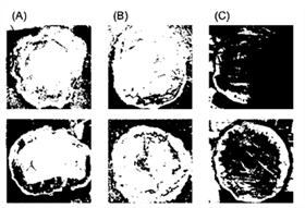
The Q. mongolica used in this study were collected from Hongcheon, Gangwon-do, Korea. The harvested timbers with decay or discoloration in the heartwood in the cross-section were selected (Fig. 1). They were transferred to the laboratory as slices and crushed into sawdust forms using a freeze-grinder (SPEXTM SamplePrep 6870 Freezer/MillTM, LLC, Metuchen, NJ, USA). During this process, samples for the isolation of fungi were prepared, and it took 1 week after harvesting.

wood-49-3-234-g1
Fig. 1. The cut surface of the deteriorated Q. mongolica.
Download Original Figure
2.2. Isolation of fungi

Two independent experiments were performed to isolate fungi among microorganisms in the sawdust.  As one experiment, 10 g of sawdust was put in the center of PD agar plate (DifcoTM potato dextrose agar, catalog number: 213400, Becton, Dickinson and Company, Franklin Lakes, NJ, USA), and the growth of microorganisms around it was monitored. As for the other experiment, 10 g of sawdust was vigorously mixed in 100 mL of 0.7% NaCl, and 10x serial dilutions were performed. 100 μL of each dilution was dispensed on PD agar plate and evenly spread using a glass spreader. PD agar plates prepared by these two methods were cultured at 25°C and a single fungus was separated depending on the growth rate.

2.3. Extraction and identification of genomic DNA from fungi

Genomic DNA was extracted from the isolated and cultured fungi from the previously described method (Umesha et al., 2016). After transferring the fungi to a 1.5 mL Eppendorf tube, they were strongly ground with a sterilized stainless steel rod. 300 μL of extraction buffer (200 mM Tris-HCl (pH 8.0), 250 mM NaCl, 25 mM EDTA (pH 8.0), 0.5% SDS) was added to them and mixed by vigorous vortex. Samples were centrifuged at 4°C, at 13,000 rpm for 15 minutes using 5417R Refrigerated Centrifuge (Product number: 5407 14618, Eppendorf Scientific, Germany). 250 μL of the supernatants were transferred to fresh Eppendorf tubes and 250 μL of phenol-chloroform-isoamyl alcohol (25:24:1 Phenol:Chloroform:Isoamyl alcohol, Catalog number: C-9017, Bioneer, Korea) was added. After gentle mixing by converting, they were centrifuged at 4°C and 13,000 rpm for 5 minutes. 500 μL of the supernatants and 500 μL of cold ethanol diluted with 95% (99.5%, Catalog number: 056-03341, Wako, Japan) were added and mixed gently. After centrifuging them at 4°C, at 13,000 rpm for 20 minutes, ethanol was removed. After 1 mL of cold 70% ethanol was added and slowly mixed, samples were centrifuged at 4°C and 13,000 rpm for 10 minutes. After ethanol was completely removed, samples were dried with a cap left opened at room temperature for 1 hour. Samples were re-suspended with 30 μL of sterilized distilled water containing 10 μg/mL RNase (RNase A Powder for K-3030, Catalog number: KB-0101, Bioneer, Korea) and reacted at 37°C for 30 minutes to obtain genomic DNA.

To amplify Internal Transcribed Spacer (ITS) region from extracted genomic DNA, AccuPower® PCR PreMix (Catalog number: K-2016, Bioneer, Korea) was used, and reaction conditions were followed by the manual. Two primers (ITS1: 5’-TCCGTAGGTGAACCTGCGG-3’, and ITS4: 5’-TCCTCCGCTTATTGATATGC-3’) were used. The amplified PCR products were purified using AccuPrep® PCR/Gel DNA Purification Kit (Catalog number: K-3037, Bioneer, Korea) according to the manual. The sequencing of purified DNA was performed at National Instrumentation Center for Environmental Management (NICEM, Seoul, Korea) using ABI 3730x1 DNA analyzer (Catalog number: A41046, Thermo Fisher Scientific Korea Ltd., Korea). Homology analyses were performed on the produced sequence using nucleotide BLAST search at National Center for Biotechnology Information (NCBI).

2.4. Phylogenetic tree analysis

Among homology search results of five fungi through nucleotide BLAST using nucleotide sequence obtained from PCR with their ITS regions as primers, the one with the highest homology was selected for each. Multiple sequence alignment for the selected nucleotide sequences was performed using ClustalW of the MEGA4 program. Neighbor-Joining (NJ) method was used for phylogenetic tree analysis of aligned nucleotide sequences, and bootstrap (1,000 replications) was applied for the reliability of the results.

2.5. Monitoring the growth of selected fungi

The pH and temperature-dependent growth of fungi was monitored in PD agar plates with 90 mm diameter. Fungi were inoculated at the center of each PD agar plate with controlled pH conditions for 4 pH values (4.5, 5.0, 5.5, and 6.0) and cultured at 3 temperature conditions (25, 28, and 30°C) for each. All experimental results were calculated using IBM® SPSS software (Ver. 25.0; SPSS Inc., Chicago, Illinois, USA) for means and standard deviations. Results were analyzed by Duncan test method of One-way analysis at the 95% confidence level. The culture period of each fungi was determined as 80% of the plate area was covered by mycelium and the diameter of the ring at this time was measured to compare the growth rate of each fungus according to the culture conditions.

Formula: Growth rate (cm/day) = mycelium diameter (cm) / culture period (day)

Where, the diameter of a mycelium and culture period are based on when about 80% of the plate is filled with fungi. Also, the average mycelium diameter formed under all culture conditions is used.

2.6. Measurement of fungal extracellular enzyme activity

Among the extracellular enzymes secreted by the fungus, the activities of cellulase and laccase were measured (Jeon and Ka, 2014). An inoculum of 6 mm diameter size was isolated and prepared from the strains cultured at 25°C on a PD agar plate. Carboxymethylcellulose sodium salt (CMC, Product number: C5678, Sigma, Korea) was selected as a substrate for the cellulase activity measurements. An inoculum with 6 mm diameter was inoculated on the center of each 50 mm diameter CMC agar (0.2% NaNO3, 0.1% K2HPO4, 0.05% MgSO4, 0.05% KCl, 0.2% carboxymethylcellulose sodium salt, 0.02% peptone, 1.5% agar, w/v) plate and cultured at 25°C in the darkness for 2 days. Then, 1-2 mL of Gram’s iodine solution (0.67% KI, 0.003% I2, w/v) was poured onto each plate and reacted at room temperature in the darkness for 2 hours. After 2 hours, the diameter of the zone showing cellulase activity was measured. 2,2’-azino-bis(3-ethylbenzthiazoline-6-sulfonic acid) diammonium salt (ABTS, Product number: A1888, Sigma, Korea) was used as a substrate for the laccase activity measurements. An inoculum with 6 mm diameter was inoculated on the center of each 90 mm diameter ABTS agar (0.2% K2HPO4, 0.2% KCI, 0.2% MgSO4·7H2O, 1.5% agar, pH 5.0, w/v) plate and cultured at 25°C in the darkness for 5 days. Five days later, the size of the turquoise blue ring formed around the inoculum was measured.

2.7. Evaluation of wood-decaying ability by fungi

The wood-decaying ability by fungi on Q. mongolica was evaluated following KS F 2213 : 2004 standard. A testing strain for broad-leaved trees, Trametes versicolor was used as a control group (Won et al., 2018). About 250 g of quartz sand dried after washing with water was filled in a 500 mL glass bottle. 80 mL of culture medium (2.5% glucose, 1% malt extract, 0.5% peptone, 0.3% KH2PO4, 0.2% MgSO4·7H2O) was evenly absorbed and sealed, and then sterilized at 121°C for 30 minutes. Heartwood and sapwood of Q. mongolica were prepared in a size of 20 x 20 x 20 mm, respectively. The prepared samples were dried at 60°C for 48 hours, then allowed to cool down in a desiccator for 30 minutes (conditioning), and their weight (W1) values were measured. Five strains of fungi were cultured for 5 days in PD (DifcoTM potato dextrose broth, catalog number: 254920, DB, USA) broth at 25°C and 150 rpm conditions, and 3 mL of culture were evenly applied to the surface of the quartz sand. Three prepared samples were put in each bottle, and then decayed at 25°C and a humidity of 70% or more, for about 40 days. The fungi adhering to the sample surface were removed thoroughly with a soft brush in flowing water and air-dried for about 20 hours. Then, they were dried at 60°C temperature condition for 48 hours, and then allowed to cool down in a desiccator for 30 minutes (conditioning), and their weight (W2) values were measured. The weight reduction rate was calculated by the formula below, and the decay ability of five fungi strains was evaluated.

Formula: Weight reduction rate (%) = (W1 – W2)/W1 x 100

W1: weight of the sample before fungal inoculation

W2: weight of the sample after fungal inoculation

3. RESULTS and DISCUSSION

3.1. Identification and phylogenetic tree analysis by nucleotide sequencing of fungi

The nucleotide sequences were obtained by PCR amplification of the ITS1 and ITS4 regions of genomic DNA extracted from the fungi. Obtained nucleotide sequences were compared to pre-existing databases using NCBI, nucleotide BLAST search. As a result (Table 1) of analyzing the nucleotide sequence homology of the fungi isolated from the decaying wood shown in Fig. 1(A), Fungal sp. was ranked at the top, followed by Umbelopsis isabellina. The nucleotide sequence of fungi isolated from the decaying wood illustrated in Fig. 1(B) confirmed them to be fungi belonging to Ophiostoma species and Ophiostoma piceae accounted for 100% in query coverage and identities. Three morphologically distinct three fungi were isolated from the decaying wood shown in Fig. 1(C). The homology comparison from one of them showed that all of the top five were identified as Aureobasidium melanogenum, and query coverage and identities were 99%. Another was confirmed to be Mucor circinelloides, and query coverage and identities were 99%. The third also showed that all of the top 5 were identified as Cunninghamella elegans, and the top result showed 99% in query coverage and 100% identities. Based on these results, the isolated fungi were suggested to be O. piceae and A. melanogenum which belong to Ascomycota phylum, and M. circinelloides, U. isabellina and C. elegans, three species that belong to Mucoromycota phylum.

Table 1. Identification results of fungi isolated from deteriorated Q. mongolica
Origin Description Max / Total score Query coverage /
Identities (%)
E-value Accession
Fig. 1(A) Fungal sp. 3.12.4D 1109 / 1109 99 / 99 0.0 KM068430.1
Umbelopsis isabellina 1083 / 1083 99 / 98 0.0 KC489502.1
Umbelopsis isabellina 1083 / 1083 99 / 98 0.0 AJ876493.1
Umbelopsis isabellina 1077 / 1077 99 / 98 0.0 KJ028792.1
Fungal endophyte sp. P1406O 1077 / 1077 98 / 99 0.0 EU977237.1
Fig. 1(B) Ophiostoma piceae 1188 / 1188 100 / 100 0.0 KF531618.1
Ophiostoma quercus 1179 / 1179 99 / 100 0.0 KM100571.1
Ophiostoma piliferum 1175 / 1175 100 / 99 0.0 KU756609.1
Ophiostoma querci 1175 / 1175 100 / 99 0.0 AF493243.1
Ophiostoma quercus 1173 / 1173 99 / 99 0.0 MH055670.1
Fig. 1(C) Aureobasidium melanogenum 1068 / 1068 99 / 99 0.0 KY659501.1
Aureobasidium melanogenum 1068 / 1068 99 / 99 0.0 KY294711.1
Aureobasidium melanogenum 1068 / 1068 99 / 99 0.0 KY294710.1
Aureobasidium melanogenum 1068 / 1068 99 / 99 0.0 KY294709.1
Aureobasidium melanogenum 1068 / 1068 99 / 99 0.0 KY294708.1
Fig. 1(C) Mucor circinelloides 1177 / 1177 99 / 99 0.0 JN561250.1
Mucor circinelloides 1170 / 1170 99 / 99 0.0 DQ118987.1
Mucor circinelloides 1168 / 1168 99 / 99 0.0 KP132465.1
Mucor circinelloides 1166 / 1166 99 / 99 0.0 KY933391.1
Mucor circinelloides 1164 / 1164 99 / 99 0.0 GU966516.1
Fig. 1(C) Cunninghamella elegans 1245 / 1245 99 / 100 0.0 JN205887.1
Cunninghamella elegans 1240 / 1240 99 / 100 0.0 JN205882.1
Cunninghamella elegans 1240 / 1240 99 / 99 0.0 FJ792589.1
Cunninghamella elegans 1234 / 1234 99 / 99 0.0 JF439470.1
Cunninghamella elegans 1232 / 1232 99 / 99 0.0 JN205884.1
Download Excel Table

Phylogenetic tree analysis was performed by selecting one each with the highest max/total score and query coverage/identities from the homology search results using the nucleotide sequences of the five fungi obtained (Fig. 2). As a result, O. piceae (KF531618.1) and A. melanogenum (KY659501.1) were grouped into one group, M. circinelloides (JN561250.1), U. isabellina (KC489502.1), and C. elegans (JN205887.1) formed another group. These two groups were separated in the early stage, and it was confirmed that the similarity between these two lines was quite distant. In one group, M. circinelloides and U. isabellina were bound to the same branch, but C. elegans initially formed different branches, meaning that C. elegans had a distance in lineage from the other two species. On the

wood-49-3-234-g2
Fig. 2. Phylogenetic analysis of five fungi nucleotide from a BLAST search at the National Center for Biotechnology Information. A neighbor-joining method with a bootstrap of 1,000 replications was used.
Download Original Figure

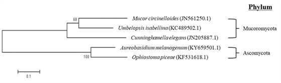
other hand, A. melanogenum and O. piceae showed a value of 100, which was considered to have high similarity in their nucleotide sequences.

3.2. Morphological characteristics of fungi isolated from deteriorated Q. mongolica

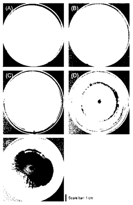
Five morphological distinct fungi were isolated and cultured from deteriorated Q. mongolica at 25°C temperature condition using PD agar as a nutrient source (Fig. 3). After observation of mycelia from 5 fungi, the surface texture of M. circinelloides and C. elegans was downy surface texture (Fig. 3(A) and 3(B)). Also, in the case of U. isabellina (Fig. 3(C)), the surface of the formed mycelium showed a rough texture. On the other hand, the mycelium of O. piceae and A. melanogenum has a relatively flat surface, smooth texture, and glossy (Fig. 3(D) and 3(E)). Also, M. circinelloides, C. elegans and U. isabellina had unclear boundaries of their mycelia, but O. piceae formed dark circular bands like annual ring around the inoculum, as the mycelium spread. In addition, A. melanogenum showed bright color in early stage mycelium but showed a tendency to darken over time forming dark brown color around the inoculum. In summarizing the morphological characteristics, it was confirmed that M. circinellodes, C. elegans, and U. isabellina formed one group, and O. piceae and A. melanogenum formed the other group. This result of being divided into two groups supported well the result of phylogenetic tree analysis using nucleotide sequences (Fig. 2), which were classified into Mucoromycota phylum and Ascomycota phylum. If the characteristics of fungi are investigated through the unique morphological analysis, it is considered that it will be useful for the initial classification of strains before a genetic identification process.

wood-49-3-234-g3
Fig. 3. Morphological characteristics of mycelial colonies isolated from deteriorated Q. mongolica. All fungi were inoculated on 90 mm diamter PD agar plate and cultured at 25°C. (A) M. circinelloides, (B) C. elegans, (C) U. isabellina, (D) O. piceae, (E) A. melanogenum.
Download Original Figure
3.3. Observation of growth characteristics of identified fungi

In order to observe the growth characteristics of the five fungi strains isolated and identified from Q. mongolica depending on the culture conditions, fungi were inoculated into PD agar plates adjusted to various pH values (pH 4.5, pH 5.0, pH. 5.5, and pH 6.0) and incubated at temperature conditions of 25°C, 28°C, and 30°C. The inoculated fungi were incubated until the surface of a plate was more than 80% covered, and the incubation period was varied according to the growth rate of each strain. Also, the diameter of their mycelia was measured to calculate the growth rate according to the cultivation period to compare the growth rate among fungi (Table 2).

Table 2. Comparisons of mycelial growth according to pH and temperature conditions
Species Mycelial growth on PD agar plate (cm)* Time**
(day)
Growth rate***
(cm/day)
pH 4.5 pH 5.0 pH 5.5 pH 6.0
Mucor circinelloides 25˚C 7.1±0.5 6.9±0.4 7.0±0.1 7.2±0.2 3 2.4
28˚C 7.2±0.2 7.3±0.2 7.7±0.6 7.5±0.3
30˚C 7.2±0.5 7.8±0.9 7.3±0.2 7.6±0.2
Cunninghamella elegans 25˚C 6.1±0.2 6.0±0.4 6.3±0.4 5.8±0.6 3 2.1
28˚C 6.9±0.4 6.7±0.5 6.8±0.0 6.2±0.2
30˚C 6.4±0.1 6.2±0.3 6.7±0.2 6.4±0.8
Umbelopsis isabellina 25˚C 7.0±0.3 7.2±0.2 7.5±0.1 7.6±0.1 9 0.8
28˚C 7.3±0.3 7.5±0.2 7.5±0.3 7.8±0.3
30˚C 6.7±0.2d,h 7.5±0.1 7.5±0.2 7.9±0.2a,i
Ophiostoma piceae 25˚C 7.5±0.2 7.0±0.8 7.4±0.7 7.0±0.1 17 0.4
28˚C 6.7±0.6 6.9±0.4 7.0±0.1 6.8±0.1
30˚C 6.1±0.1 6.1±0.3 5.7±0.1a 5.9±0.2a
Aureobasidium melanogenum 25˚C 7.5±0.3 7.1±0.1 7.5±0.1 7.3±0.1 14 0.5
28˚C 7.4±0.2 7.3±0.1 7.2±0.1 7.0±0.2
30˚C 6.6±0.1a,c,d,e,f 6.7±0.2a,c 6.6±0.2a 6.4±0.3a,b,c,d,e,f,g

* The sizes of mycelium formed under various culture conditions were compared for each fungi at a 95% confidence level.

** The day when 80% of the surface of the plate is covered.

*** Average fungi growth rate under all culture conditions.

ap < 0.05 vs. 25°C, pH 4.5; bp < 0.05 vs. 25°C, pH 5.0; cp < 0.05 vs. 25°C, pH 5.5; dp < 0.05 vs. 25°C, pH 6.0; ep < 0.05 vs. 28°C, pH 4.5; fp < 0.05 vs. 28°C, pH 5.0; gp < 0.05 vs. 28°C, pH 5.5; hp < 0.05 vs. 28°C, pH 6.0; ip < 0.05 vs. 30°C, pH 4.5

Download Excel Table

After checking the growth rate for each species, it took 3 days until 80% of the plate surface was covered with mycelium by M. circinellodes and C. elegans, and the growth rate were confirmed to be 2.4 cm/day and 2.1 cm/day, respectively. Also, U. isabellina was required 9 days of the culture period, and its growth rate was confirmed to be 0.8 cm/day. O. piceae and A. melanogenum needed 17 days and 14 days to form sufficient mycelia, respectively, and the determined growth rate was confirmed to be 0.4 cm/day and 0.5 cm/day. From these results, the growth rates of M. circinelloides, C. elegans, and U. isabellina belonging to Mucoromycota phylum isolated and identified through this study were found to be 2 – 6 times faster than those of O. piceae and A. melanogenum belonging to Ascomycota phylum.

Growth changes depending on pH and temperature conditions were examined for each species, and basic physiological characteristics of each strain were investigated by observing a significant difference in growth with respect to the size of the mycelium measured under each condition. As a result, no significant pH/temperature dependence was observed from M. circinelloides and C. elegans. In contrast, U. isabellina formed a wider mycelium cultured at pH 6.0 than pH 4.5, at both 25°C and 30°C culture temperatures (p < 0.05). Based on these results, it is suggested that pH is a more important factor for the growth of U. isabellina than temperature. In the case of O. piceae, a wider mycelium was formed when cultured at pH 5.5 and 25°C than pH 5.5 and 30°C within the same period (p < 0.05). It is considered that relatively low temperature is suitable for the formation of O. piceae mycelium. The growth of A. melanogenum generally formed a wider mycelium when cultured at 25°C than 30°C (p < 0.05). From these results, it is suggested that relatively low temperatures are suitable for the growth of A. melanogenum.

Despite some differences in the culture period, all strains showed active growth enough to cover all the plates surface regardless of the culture conditions. This phenomenon is suggested that glucose, which is the main constituent of PD agar used for culture in this study, is the preferred energy source by most microorganisms, and therefore optimal culture conditions have not been derived. It is considered that further investigations will be necessary to observe the nutrient sources favored by the five fungi and the morphological characteristics expressed at that moment, by varying the medium conditions depending on carbon and nitrogen sources.

3.4. Extracellular enzyme activity of fungi

Woods have complex layered structures composed of polymer compounds of cellulose, hemicellulose, and lignin. Wood decaying fungi and other wood-harming microorganisms must have a system which is capable of decomposing these chemical structures to effectively decompose woods and obtain nutrients (Bidlack et al., 1992Brändström, 2001; Yoon et al., 2018). Since the five fungi strains obtained in this study were isolated from deteriorated Q. mongolica, with decay and discoloration, it was expected that they would be able to decompose wood biomass. So, the presence or absence of enzymes that degrade cellulose and lignin were confirmed by using CMC and ABTS as substrates, respectively.

CMC is one of the cellulose derivatives (Hankin and Anagnostakis, 1977), and its β-1,4-glucosidic bond is hydrolyzed by cellulase to form glucose (Percival Zhang et al., 2006; Yoon et al., 2019). By inoculating 5 types of fungi on a 5.0 cm diameter agar plate containing CMC and incubating at 25°C, a ring was formed by cellulase activity around all inocula (Fig. 4(A)). Especially, M. circinelloides and C. elegans formed the biggest rings with the size of 4.3 and 4.0 cm, respectively. In contrast, U. isabellina, O. piceae, and A. melanogenum formed relatively small rings, 2.0, 1.9, and 1.8 cm, respectively. M. circinelloides and C. elegans, which formed big rings, belong to Mucoromycota phylum, and O. piceae and A. melanogenum, which formed relatively small rings, are classified as Ascomycota phylum. Laccase is an extracellular enzyme produced by most fungi and has been reported to break down lignin in wood (Baldrian, 2006; Giardina et al., 2010). By confirming the secretion of laccase, a lignin-degrading (ligninolytic) enzyme, with ABTS assay, the possibility of lignin decomposition capability of five fungi strains was confirmed. After inoculating strains on the agar plate containing ABTS and incubating for 5 days, the secretion of laccase was confirmed by the presence or absence of a turquoise ring (Fig. 4(B)). As a results, turquoise rings of 3.2 cm and 2.1 cm were formed around the inoculum of O. piceae and A. melanogenum, respectively. On the other hand, M. circienlloides, C. elegans, and U. isabellina did not form a ring induced by laccase decomposition. Interestingly, the two fungi strains, O. piceae and A. melanogenum, which their laccase secretion was confirmed by the formation of turquoise rings, belong to Ascomycota phylum, and the other three strains that do not secrete laccase belong to Mucoromycota phylum.

wood-49-3-234-g4
Fig. 4. Cellulase (A) and laccase (B) activities of five fungi isolated in this study. The cellulase and laccase activities of fungi were measured using CMC and ABTS, respectively, as substrates. All culture conditions were 25°C, and each activity was measured after two days for cellulase and five days for laccase. CMC assay was performed on a 50 mm plate, and ABTS assay was performed on a 90 mm plate. A. M. circinelloides, B. C. elegans, C. U. isabellina, D. O. piceae, E. A. melanogenum. N.D is an abbreviation for not detected, meaning that no enzyme activity was observed.
Download Original Figure

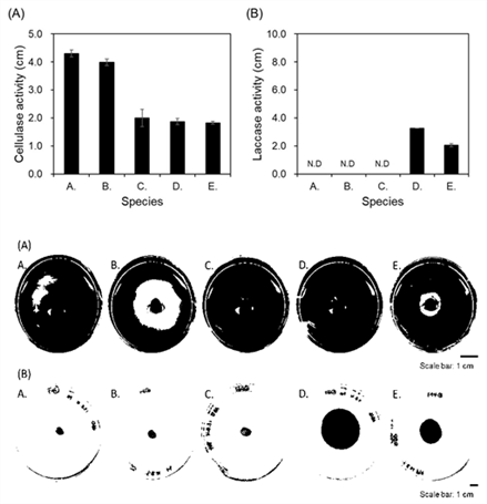
M. circinelloides, which is commonly found in soil, plants, and decayed fruits (Carvalho et al., 2015; Lee et al., 2014), has the nucleotide sequence of β-glucosidase, a type of cellulase, and it has been reported to show enzymatic activities (Huang et al., 2014). The M. circinelloides strain isolated and identified in this study also exhibited cellulase activity, and it is presumed to be a fungus involved with the deterioration of Q. mongolica. C. elegans has lignin peroxidase enzyme, and it has been reported to show the maximum enzyme activity at 30°C temperature and pH 6.0 conditions (Roushdy et al., 2011). However, the C. elegans strain obtained in this study did not show ligninolytic activity, and it might be explained to be caused by the difference in conditions for measuring the enzyme activity. Thus, it was considered that it is necessary to perform different temperature and pH conditions in the screening process to evaluate the presence or absence of enzyme activity as well. O. piceae, in which cellulase and laccase activities were observed, is a soft-rot fungus and has been reported to cause discoloration as well as decay of woods (Abraham et al., 2011). On the other hand, U. isabellina and A. melanogenum have not been reported on wood-degrading enzymes. However, in this study, cellulase activity of U. isabellina and cellulase and laccase activity of A. melanogenum were observed, so they would be related to the wood decay. Based on these results, among 5 fungi strains isolated from Q. mongolica in this study, it is expected that O. piceae and A. melanogenum, which belong to Ascomycota phylum, secrete both cellulase and laccase enzymes, involving with degrading cellulose and lignin of Q. mongolica.

In addition, it is considered that M. circinellodes, C. elegans, and U. isabellina, belonging to the Mucoro-mycota phylum, secrete cellulase and affect cellulose decomposition in Q. mongolica.

3.5. Evaluation of the decaying ability of fungi on Q. mongolica

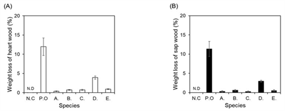
All of the five fungi isolated from the deteriorated Q. mongolica possessed cellulase activity. O. piceae and A. melanogenum among them showed laccase activity as well as cellulase activity. Based on these results, the 5 fungi identified in this study are expected to be related to the deterioration of Q. mongolica, so their decaying ability was experimentally examined on Q. mongolica. Because fungi with enzyme activity decompose the constituents of wood and use them as a nutrient source, the weight of woods decayed by them should be reduced. Therefore, the weight reduction rates of five strains of fungi were measured for heartwood and sapwood of Q. mongolica, and used as a criterion for evaluating the wood-decaying ability (Fig. 5). Results showed that the weight reduction rate of Q. mongolica heartwood by O. piceae was about 3.9% and sapwood was about 3.0%. On the other hand, the rest of the strains had insignificant effects on weight reduction rate which were showing less than 1% reduction for both heartwood and sapwood. When comparing the weight reduction rate by each fungus for the heartwood and sapwood, O. piceae and A. melanogenum, belonging to Ascomycota phylum, showed 6.9% and 1.4%, respectively, which were relatively higher than strains belonging Mucoromycota phylum: M. circinelloides (0.7%), C. elegans (1.3%), and U. isabellina (1.0%). Fungi belonging to Ascomycota phylum are also called soft-rot fungi, and they are found in both softwoods and hardwoods, but found more in hardwoods and are known to cause more damages. It has been reported that wood contaminated with soft-rot fungi undergo damages including softening surface and discoloration to dark brown (Nilsson and Daniel, 1989; Song et al., 2010).

wood-49-3-234-g5
Fig. 5. Deterioration test on heartwood (A) and sapwood (B) of five fungi isolated in this study. The wood decay test was carried out for forty days at 25°C and 70% humidity conditions. N.C: negative control (No fungi inoculation) P.O: positive control (Trametes versicolor) A. M. circinelloides, B. C. elegans, C. U. isabellina, D. O. piceae, E. A. melanogenum. N.D is an abbreviation for not detected, meaning that no weight loss was observed.
Download Original Figure

As previously described, all 5 fungi strains showed cellulase activity confirmed by CMC assay and ABTS assay, and two fungi belonging to Ascomycota phylum were found to possess laccase activity as well as cellulase activity. Lignin surrounds cellulose, and hemicellulose acts as a bridge between the two bonds (Carrier et al., 2017; Zhang et al., 2015). This tightly bonded lignin maintains the shape of a wood or protects cellulose from external invasion (Xie et al., 2018). So, the decomposition of lignin by the laccase enzyme activity helps the decaying microorganisms with enzymatic activity including cellulase to penetrate into the wood, suggesting the possibility that the decay of woods can proceed more effectively. Therefore, it is considered that the decomposition of lignin by laccase activity as well as cellulase activity acts as an important factor in the decaying of Q. mongolica, and it is suggested that O. piceae and A. melanogenum, belonging to Ascomycota phylum, which possess both enzyme activities, play a more important role in it.

4. CONCLUSION

From this study, overall five fungi strains were isolated and identified from Q. mongolica with discoloration, decay, and deterioration damages. The identified fungi were confirmed to be three species that belong to Mucoromycota phylum including M. circinelloidesC. elegans and U. isabellina, and two species that belong to Ascomytoca phylum including O. piceae and A. melanogenum. By evaluating the enzyme activity that affects wood decay, cellulase activity involved in cellulose degradation was observed in all fungi. Also, unlike the other fungi, O. piceae and A. melanogenum showed cellulase activity as well as laccase activity that degrades lignin. The weight reduction effect was also observed by all five fungi in the evaluation of wood decaying performed on the heartwood and sapwood of Q. mongolica. Based on these results, it is considered that the five fungi isolated from the deterioration of Q. mongolica have enzymes that degrade cellulose and lignin, which are major components of wood, and thus can affect wood decaying. Therefore, the identified fungi are considered to be directly involved with the decay of Q. mongolica, but in order to control the decay, further studies on the pathways and mechanisms by which these fungi are infected in living Q. monoglica will be needed.

Appendices

APPENDIX(Korean Version)
신갈나무의 부후에 관여하는 곰팡이 분리 및 동정

초록: 신갈나무는 국내 전역에 두루 분포되어 있는 경제, 산업적으로 활용 가치가 큰 수종이지만, 변색, 부후 등의 열화에 의한 피해가 심각하다. 이러한 이유로 신갈나무의 부후는 목재로써의 활용에 걸림돌이 되나, 부후 요인에 대한 연구는 미비한 실정이다. 본 연구에서는 신갈나무 부후에 영향하는 요인으로 곰팡이에 주목하였으며, 신갈나무의 부후 부위로부터 곰팡이를 분리, 동정하였다. 또한, 동정 된 곰팡이가 실제로 목재 열화에 영향을 미치는지 확인하기 위해 효소 활성을 평가하고, 곰팡이를 처리한 신갈나무 목재의 질량 손실을 목재 부후 실험을 통해 측정하였다. 신갈나무에서 분리된 곰팡이 5종의 genomic DNA의 ITS region을 이용한 염기서열 분석을 통해, Mucoromycota phylum에 속하는 Mucor circinelloides, Cunninghamella elegans, 그리고 Umbelopsis isabellina 3종과 Ascomycota phylum에 속하는 Ophiostoma piceaeAureobasidium melanogenum 2종의 곰팡이가 동정되었다. 이러한 5종의 곰팡이는 목재의 부후와 관련된 cellulase나 laccase와 같은 효소 활성이 있으며, 실제로 신갈나무의 심재와 변재의 중량을 감소시켰다. 특히, cellulase와 laccase 활성을 모두 보유한 O. piceaeA. melanogenum는 신갈나무의 중량을 각각 6.9%와 1.5% 감소시켰다. 이러한 결과들은 본 연구에서 동정된 5종의 곰팡이가 신갈나무의 열화에 영향한다는 것을 의미하며, 신갈나무에 대한 목재 부후균으로써의 가능성을 시사한다.

1. 서론

낙엽 활엽 교목인 참나무속은 산림생태계에 중요한 생태 서비스를 제공하며(Plomion and Fievet, 2013), 인간에게 중요한 목재 및 생체화합물 등의 자원을 공급한다(Li et al., 2010; Chang et al., 2017). 그 중, 신갈나무(Quercus mongolica)는 국내 총 1,372종의 참나무류 중에서 308본으로 약 28.4%의 비율을 차지하는 우점종으로, 우리나라의 전역에 두루 분포되어 있으며(Lam et al., 2015; Lee et al., 2018), 그 면적은 229,821 ha에 이르는 것으로 보고된 바 있다(Korea Forest Service, 2018). 신갈나무의 열매는 식용이 가능하고, 그 목재는 건축재, 기구재 등의 원료로써 경제, 산업적으로 사용되며(Son et al., 2004), 항균, 항염증, 항산화 등의 생리 활성 물질이 함유되어 있는 등 활용가치가 높은 수종으로 알려져 있다(Eo et al., 2019; Kong et al., 2001; Yin et al., 2019; Jung et al., 2017; Jung et al., 2017).

그러나, 신갈나무는 수피의 코르크층 발달이 상대적으로 적고, 맹아 발생률이 높아 외부의 상처나 미생물의 침투에 쉽게 노출되어, 부후에 의한 고사율이 높은 수종으로 보고되었다(Lee et al., 2017; Jeon et al., 2020). 선행 연구 보고에 따르면 전국에서 벌채한 2,504본의 신갈나무의 부후율이 66.1%에 이르는 것으로 조사되었으며, 대부분이 조직고사 및 변색 단계의 부후 형태를 나타내는 것으로 확인되었다. 게다가 직경과 임령이 증가할수록 부후율이 증가하는 것으로 보고되었다(Kang et al., 2020). 부후가 진행된 신갈나무는 내부의 재색이 갈색이나 흑갈색으로 변색되거나, 공동이 형성되는 등의 결함이 발생되어 품질 등급이 하락하기 때문에 심각한 경제적 피해가 야기된다. 이처럼 신갈나무의 부후는 경제적 이용에 장애가 되고 있으나, 부후에 대한 명확한 원인 구명이 이루어져 있지 않다. 신갈나무의 고부가가치화를 위해서는 이러한 현상에 대한 원인 구명을 통해 신갈나무 부후를 제어할 수 있는 방안마련이 필요한 실정이다.

부후를 비롯한 목재의 열화(deterioration of wood)는 물리, 화학 특성이 분해되거나 변질되는 현상이며, 일반적으로 미생물이나 벌레에 의한 생물 열화와 이 밖의 원인에 의해 발생되는 비생물 열화의 두 가지 종류로 크게 구분된다(Brischke et al., 2006; Brischke and Rapp, 2010; Teodorescu et al., 2017). 목재의 줄기, 가지 또는 뿌리를 약화시키는 부후의 피해를 야기하거나 변색을 유발하는 등 생물 열화에 관여하는 요인으로 목재부후균인 곰팡이가 대표적이다(Brischke et al., 2006; White, 2004). 곰팡이에 의한 목재의 부후는 심, 변재의 고유한 색이나 강도를 변질시키며, 이러한 목재는 이용 가치가 떨어지기 때문에 경제적으로 손실이 발생한다(Schwarze, 2007). 일반적으로 자연 상태에서의 생물 열화는 주변 환경 조건에 따라 다양한 곰팡이들이 관여하여 복잡한 과정으로 진행된다. 따라서, 부후에 관여하는 곰팡이들은 수목의 종류에 따라 다르게 조사될 뿐만 아니라, 세포 외로 분비하는 cellulase나 laccase와 같은 분해 효소의 활성의 정도 또한 상이하기 때문에 특정 수목마다 부후에 관여하는 곰팡이의 연구가 필요하다(Clausen, 1996; Hildén and Mäkelä, 2018; White, 2004).

앞서 국내와 일본에서는 경제 수종인 낙엽송에 대하여 부후균에 의한 피해를 예방하고 진단하기 위해 부후에 관여하는 곰팡이의 연구가 보고된 바 있다(Kim, 1996; Kim, 1997). 부후 피해를 입고 있는 낙엽송과 같이 신갈나무의 부후에도 미생물이 관여되어 있을 것으로 판단된다. 따라서 본 연구에서는 신갈나무의 부후, 변색 등 열화에 관여하는 곰팡이를 확인하고자 신갈나무 부후 부위로부터 곰팡이를 분리 및 동정하였다. 또한 동정된 곰팡이의 cellulase, laccase 효소활성, 목재 부후력을 평가함으로써 신갈나무의 부후에 직접적인 영향을 줄 수 있는지를 확인하였다.

2. 재료 및 방법
2.1. 공시재료

본 연구에 사용된 신갈나무는 강원도 홍천에서 채취하였다. 벌채 되었을 때 절단면의 심재 부위가 변색 또는 부후된 목재를 선별하였다(Fig. 1). 벌채된 목재는 절편으로 제작되어 연구소로 운송되었으며, 동결 분쇄기(SPEXTM SamplePrep 6870 Freezer/MillTM, LLC, Metuchen, NJ, USA)를 이용하여 톱밥 형태로 분쇄되었다. 이러한 과정을 거쳐 곰팡이 분리를 위한 시료가 준비되었으며, 벌채로부터 총 일주일의 기간이 소요되었다.

2.2. 곰팡이 분리

톱밥에 존재하는 미생물 중, 곰팡이를 분리하기 위해서 두 가지 독립적인 실험을 수행하였다. 하나의 실험은 톱밥 10 g을 PD agar(DifcoTM potato dextrose agar, catalog number: 213400, Becton, Dickinson and Company, Franklin Lakes, NJ, USA) plate의 중앙에 놓고 주변으로 자라는 미생물을 관찰하였다. 다른 하나의 실험은 우선, 톱밥 10 g을 100 mL의 0.7% NaCl와 강하게 혼합한 후, 이를 10배씩 연속 희석하였다. 각 희석물들을 PD agar plate에 100 μL씩 분주하고, glass spreader를 이용하여 고르게 도포하였다. 두 가지 방법으로 준비된 PD agar plate는 25°C 온도 조건에서 배양하였으며, 생장 속도에 따라서 각각의 단일 곰팡이로 분리하였다.

2.3. 곰팡이의 genomic DNA 추출 및 동정

선행 방법을 참고하여 분리 배양된 곰팡이들로부터 genomic DNA를 추출하였다(Umesha et al., 2016). 1.5 mL Eppendorf tube에 곰팡이를 옮긴 후, 멸균된 스테인리스 막대로 강하게 갈아주었다. 300 μL의 extraction buffer (200 mM Tris-HCl (pH8.0), 250 mM NaCl, 25 mM EDTA (pH 8.0), 0.5% SDS)를 첨가하여 강하게 vortex하여 혼합하였다. 원심분리기(5417R Refrigerated Centrifuge, Product number: 5407 14618, Eppendorf Scientific, Germany)를 이용하여 4°C, 13,000 rpm 조건으로 15분 동안 원심분리하였다. 새로운 e-tube에 상층액 250 μL을 옮겨담고, 250 μL phenol-chloroform-isoamyl alcohol (25:24:1 Phenol:Chloroform:Isoamyl alcohol, Catalog number: C-9017, Bioneer, Korea)를 첨가하였다. 부드럽게 converting하여 혼합한 후, 4°C, 13,000 rpm 조건으로 5분 동안 원심분리하였다. 상층액 500 μL와 95%로 희석된 차가운 ethanol (99.5%, Catalog number: 056-03341, Wako, Japan) 500 μL를 첨가하여, 부드럽게 혼합하였다. 4°C, 13,000 rpm 조건으로 20분 동안 원심분리한 후, ethanol을 제거하였다. 1 mL의 차가운 70% ethanol을 첨가하여 부드럽게 혼합한 후, 4°C, 13,000 rpm 조건으로 10분 동안 원심분리하였다. Ethanol을 완벽하게 제거한 후, 뚜껑을 연 상태로 상온에서 1시간 동안 건조시켰다. 10 μg/mL RNase (RNase A Powder for K-3030, Catalog number: KB-0101, Bioneer, Korea)가 포함된 멸균된 증류수 30 μL로 resuspend하여 37°C에서 30분 동안 반응하여 genomic DNA를 확보하였다.

추출된 genomic DNA로부터 Internal Transcribed Spacer (ITS) region을 증폭하기 위해 AccuPower® PCR PreMix (Catalog number: K-2016, Bioneer, Korea)을 이용하였으며, 반응 조건은 매뉴얼을 따랐다. 이 때 두개의 primer (ITS1: 5’-TCCGTAGGTGAACCTGCGG-3’, ITS4: 5’-TCCTCCGCTTATTGATATGC-3’)를 이용하였다. 증폭된 PCR product는 AccuPrep® PCR/Gel DNA Purification Kit (Catalog number: K-3037, Bioneer, Korea)의 매뉴얼을 따라 정제하였다. 정제된 DNA product의 염기서열 분석은 National Instrumentation Center for Environmental Management (NICEM, Seoul, Korea)에서 수행하였으며, ABI 3730x1 DNA analyzer (Catalog number: A41046, Thermo Fisher Scientific Korea Ltd., Korea)를 이용하였다. 분석된 염기서열을 이용하여 National Center for Biotechnology Information (NCBI)의 nucleotide BLAST search를 통해 상동성 분석을 수행하였다.

2.4. 계통수 분석 (Phylogenetic tree analysis)

5종 곰팡이의 ITS region을 primer로 하여 PCR을 통해 얻어진 염기 서열의 nucleotide BLAST를 통한 homology search 결과 중에서, 각각 상동성이 가장 높은 1개씩을 선별하였다. 선별된 염기 서열을 MEGA4 프로그램의 ClustalW를 이용하여 multiple sequence alignment를 실시하였다. Align된 염기 서열들의 계통수 분석을 위해 Neighbor-Joining (NJ) 방법을 사용하였으며, 결과의 신뢰성을 위해 bootstrap (1,000 replications)을 적용하였다.

2.5. 선별된 곰팡이의 생장 관찰

90 mm 직경의 PD agar plate에서 온도와 pH에 따른 각 곰팡이의 생장을 관찰하였다. 4종류(4.5, 5.0, 5.5, 6.0)의 pH로 조절된 PD agar plate의 중앙에 6 mm 직경의 접종원을 하나씩 접종하고, 3종류(25, 28, 30˚C)의 온도 조건으로 배양하였다. 모든 실험 결과는 평균과 표준 편차를 계산하기 위해서 IBM® SPSS software (Ver. 25.0; SPSS Inc., Chicago, Illinois, USA)를 이용하여 계산되었다. 결과는 일원배치 분산분석(One-way analysis)의 Duncan 검정 방법으로 수행되었으며, 95% 신뢰 수준에서 정의되었다. Plate의 80% 정도가 균체로 채워졌을 때를 각 균의 배양 기간으로 하고, 이 때의 환의 직경을 측정하여 배양 조건에 따른 각 곰팡이의 생장률을 비교하였다.

계산식: 생장률 (cm/day) = 균체의 직경 (cm) / 배양 기간 (day)

단, 균체의 직경과 배양 기간은 plate의 80% 정도가 균체로 채워졌을 때를 기준으로 한다. 또한, 균체의 직경은 모든 배양 조건에서 형성된 균체의 평균값을 사용한다.

2.6. 곰팡이의 균체외 효소 활성 측정

곰팡이가 분비하는 세포 외 효소 중 cellulase와 laccase의 활성을 측정하였다(Jeon and Ka, 2014). PD agar plate에서 25°C 온도 조건으로 배양된 균주로부터 6 mm 직경의 접종원을 분리하여 준비하였다. Cellulase의 활성을 측정하기 위해 carboxymethylcellulose sodium salt (CMC, Product number: C5678, Sigma, Korea)를 기질로 선택하였다. 50 mm 직경의 CMC agar (0.2% NaNO3, 0.1% K2HPO4, 0.05% MgSO4, 0.05% KCl, 0.2% carboxymethylcellulose sodium salt, 0.02% peptone, 1.5% agar, w/v) plate의 중앙에 6 mm 직경의 접종원을 1개씩 접종한 후, 빛이 차단된 25°C 배양기에서 이틀 동안 배양하였다. 그 후, plate의 중앙에 Gram’s iodine solution (0.67% KI, 0.003% I2, w/v)을 1 – 2 mL씩 떨어뜨린 후, 알루미늄 호일로 빛을 차단하여 2시간 동안 상온에서 반응하였다. 2시간 후 cellulase의 활성이 나타나는 zone의 직경을 측정하였다. Laccase의 활성은 2,2’-azino-bis(3-ethylbenzthiazoline-6-sulfonic acid) diammonium salt (ABTS, Product number: A1888, Sigma, Korea)을 기질로 사용하여 측정하였다. 준비된 6 mm 직경의 곰팡이 접종원을 90 mm 직경의 ABTS agar (0.2% K2HPO4, 0.2% KCI, 0.2% MgSO4·7H2O, 1.5% agar, pH 5.0, w/v) plate의 중앙에 접종하여 빛이 차단된 상태로 25°C에서 5일 동안 배양하였다. 5일 후 접종원을 중심으로 형성되는 청록색의 환의 크기를 측정하였다.

2.7. 곰팡이에 의한 목재 부후력 시험

KS F 2213 : 2004 규격에 따라 신갈나무에 대한 곰팡이의 부후력을 평가하였다. 대조군은 활엽수용 공시 균인 Trametes versicolor를 사용하였다(Won et al., 2018). 500 mL 부피의 glass bottle에 물에 씻어서 건조된 석영모래를 약 250 g 채웠다. 80 mL의 배양액(2.5% glucose, 1% malt extract, 0.5% peptone, 0.3% KH2PO4, 0.2% MgSO4·7H2O)을 고르게 흡수시키고 밀봉한 후, 121°C에서 30분간 멸균하였다. 신갈나무의 변재와 심재를 각각 20 x 20 x 20 mm 크기로 제작하였다. 제작된 시료는 60°C 온도 조건에서 48시간 동안 건조한 후, 데시게이터에서 30분 동안 방냉하여(conditioning) 무게(W1)를 측정하였다. 5종의 곰팡이는 PD (DifcoTM potato dextrose broth, catalog number: 254920, DB, USA) broth에서 25°C 온도, 150 rpm 조건으로 5일 동안 배양하였으며, 석영모래의 표면에 3 mL씩 고르게 도포하였다. 준비된 시료를 각 bottle당 3개씩 넣은 후, 25°C 온도, 70% 이상 습도 조건에서 약 40일간 두어 부후시켰다. 시료의 표면에 부착된 균체를 흐르는 물에서 부드러운 브러시를 이용하여 깨끗이 닦아내고, 약 20시간 동안 바람에 건조시켰다. 그 후, 60°C 온도 조건에서 48시간 동안 건조시키고, 데시게이터에서 30분 동안 방냉하여(conditioning) 무게(W2)를 측정하였다. 시료의 중량 감소율은 다음의 계산식에 따라 계산되었으며, 이를 기준으로 5종 곰팡이의 부후력이 평가되었다.

계산식: 중량 감소율 (%) = (W1 – W2) / W1 ⅹ 100

W1: 곰팡이 접종 전 시료의 무게

W2: 곰팡이 접종 후 시료의 무게

3. 결과 및 고찰
3.1. 곰팡이의 염기 서열 분석을 통한 동정 및 계통수 분석

곰팡이로부터 추출된 genomic DNA의 ITS1과 ITS4 region을 PCR 증폭을 통해 염기 서열이 확보되었다. 확보된 염기 서열은 NCBI의 nucleotide BLAST search를 이용하여 기존의 database와 비교되었다. Fig. 1(A)의 부후 목재로부터 분리된 곰팡이의 염기 서열 상동성을 분석한 결과(Table 1), Fungal sp. 가 가장 상위에 위치하였고, 그 다음으로는 Umbelopsis isabellina들이 확인되었다. Fig. 1(B)의 부후된 목재로부터 분리된 곰팡이의 염기 서열은 Ophiostoma 종에 속하는 곰팡이들이 확인되었으며, Ophiostoma piceae가 query coverage와 identities가 각각 100%로 나타났다. Fig. 1(C)의 부후된 목재에서는 형태학적으로 구분되는 3가지의 곰팡이가 분리되었다. 그 중 하나의 상동성 비교 결과, 상위 5개가 모두 Aureobasidium melanogenum으로 확인되었으며, query coverage와 identities가 99%를 나타냈다. 또 다른 곰팡이는 모두 Mucor circinelloides가 확인되었으며, query coverage와 identities가 99%를 나타냈다. 나머지 하나도 또한, 상위 5개가 모두 Cunninghamella elegans로 확인되었으며, 가장 상위에 위치한 결과의 query coverage는 99%, identities는 100%를 나타냈다. 이러한 결과를 바탕으로, 분리된 곰팡이들은 Ascomycota phylum에 속하는 O. piceaeA. melanogenum, 그리고 Mucoromycota phylum에 속하는 M. circinelloides, U. isabellinaC. elegans 3종으로 추정된다.

확보된 5종 곰팡이의 염기 서열을 이용한 homology search 결과에서 max / total score와 query coverage / identities 수치가 가장 높은 것을 각각 1개씩 선정하여 계통수 분석을 수행하였다(Fig. 2). 그 결과, O. piceae (KF531618.1)와 A. melanogenum (KY659501.1)이 하나의 그룹으로 묶였으며, M. circinelloides (JN561250.1), U. isabellina (KC489502.1), 그리고 C. elegans (JN205887.1)이 또 다른 그룹을 형성하였다. 이 두 그룹은 초기에 분리된 것으로 계통 간의 유사도가 먼 것으로 확인되었다. 하나의 그룹에서 M. circinelloidesU. isabellina는 같은 가지로 묶여 있으나, C. elegans는 초기에 다른 가지를 형성하였으며, 이는 C. elegans가 두 종과 계통 간의 거리가 있다는 것을 의미한다. 반면에, A. melanogenumO. piceae는 100의 수치를 나타냄으로써 염기 서열 간의 유사도가 높은 것으로 사료된다.

3.2. 열화된 신갈나무로부터 분리된 곰팡이의 형태학적 특징

PD agar를 영양원으로 사용하고, 25°C의 온도 조건에서 열화된 신갈나무로부터 형태학적으로 구분되는 5종의 서로 다른 곰팡이가 분리 배양되었다(Fig. 3). 5종 곰팡이의 균사체를 관찰한 결과, M. circinelloidesC. elegans는 표면 질감이 솜털과 같았으며(Fig. 3(A)와 3(B)), U. isabellina (Fig. 3(C)) 또한 형성된 균사체의 표면이 거친 질감을 나타냈다. 반면에 O. piceaeA. melanogenum (Fig. 3(D)와 3(E))는 상대적으로 표면이 평탄하며 질감이 매끈하고 광택이 있다. 또한, M. circinelloides, C. elegans, 그리고 U. isabellina는 균사체가 퍼져나가는 경계가 뚜렷하지 않지만, O. piceae는 균사체가 퍼져나갈수록 접종원을 중심으로 나이테와 같이 짙은 색의 원형의 띠가 형성되는 것이 관찰되었다. A. melanogenum 또한, 새로 형성되는 균사체는 옅은 색을 나타냈지만, 시간이 지남에 따라 짙어지는 경향을 나타내어 접종원을 중심으로 어두운 갈색을 나타냈다. 형태학적인 결과를 바탕으로 종합했을 때, M. circinellodes, C. elegans, 그리고 U. isabellina가 하나의 그룹, 그리고 O. piceaeA. melanogenum이 또 하나의 그룹처럼 분류되는 것이 확인되었다. 이는 앞서 염기 서열을 이용한 계통수 분석 결과(Fig. 2)와 동일하게 Mucoromycota phylum과 Ascomycota phylum 두 그룹으로 구분되는 결과이다. 이처럼 곰팡들이의 고유한 형태학적 분석을 통해 그 특징을 조사한다면 유전적 동정 과정에 앞서서 일차적으로 균주의 분류 작업에 도움이 될 것이라 사료된다.

3.3. 동정된 곰팡이의 생장특성 관찰

신갈나무 부후 부위에서 분리 동정된 5종 곰팡이의 배양 조건에 따른 생장 특성을 관찰하기 위해 다양한 pH 값으로 조절된 PD agar plate (pH 4.5, pH 5.0, pH. 5.5, pH 6.0)에 접종하여, 각각 25°C, 28°C, 30°C의 온도 조건에서 배양하였다. 접종된 곰팡이로 plate의 표면이 80% 이상 덮힐 때까지 배양하였으며, 각각의 생장 속도에 따라 배양 기간을 다르게 하였다. 또한, 이 때 균사체의 직경을 측정하여 배양 기간에 따른 생장률을 계산하여 곰팡이 간의 생장률을 비교하고자 하였다(Table 2).

각 균종별 생장률을 확인한 결과, M. circinellodesC. elegans로 인해 plate의 80%가 균사체로 덮히기까지 소요된 배양 기간은 3일이었으며, 균사체 직경 대비 생장률을 계산한 결과 각각 2.4 cm/day와 2.1 cm/day로 확인되었다. 또한, U. isabellina의 경우 9일의 배양 기간이 요구되었으며, 생장률은 0.8 cm/day로 확인되었다. O. piceaeA. melanogenum은 각각 17일과 14일을 배양하였을 때 충분한 균사체의 형성을 나타냈으며, 이로부터 계산된 생장률은 각각 0.4 cm/day와 0.5 cm/day로 확인되었다. 이러한 결과로 미루어 보았을 때, 본 연구를 통해 분리 동정된 Mucoromycota phylum에 속하는 M. circinelloides, C. elegans, 그리고 U. isabellina의 생장률은 상대적으로 Ascomycota phylum에 속하는 O. piceaeA. melanogenum보다 약 2 - 6배 빠른 것으로 확인되었다.

각 균종별로 pH와 온도 조건에 따른 생장 변화를 측정하였으며, 각각의 조건에서 측정된 균사체의 크기에 대하여 유의미한 생장 차이를 관찰함으로써 각 균주들의 기본 생리 특성을 파악하고자 하였다. 그 결과, M. circinelloidesC. elegans는 각각 pH와 온도의 변화에 따른 유의미한 생장의 변화가 관찰되지 않았다. 반면, U. isabellina는 25°C와 30°C 온도의 pH 4.5 배양 조건 하에서 형성되는 균사체의 크기보다 동일 온도의 pH 6.0 조건에서 배양된 균사체가 더 넓게 형성되었다(p < 0.05). 이러한 현상으로 미루어 보았을 때, U. isabellina의 생장에는 온도보다 pH가 중요한 요인으로 작용하는 것으로 사료된다. O. piceae의 경우 25°C 온도의 pH 5.5에서 배양되었을 때, 30°C 온도의 pH 5.5 조건보다 동일 기간내에 더 넓은 균사체를 형성하였다 (p < 0.05). 이는 O. piceae 균사체 형성에는 상대적으로 낮은 온도가 적합한 것으로 사료된다. A. melanogenum의 생장은, 전반적으로 30°C에서 배양 되었을 때보다, 25°C에서 배양 되었을 때 넓은 균사체를 형성하였다(p < 0.05). 이러한 결과로 미루어 보았을 때, A. melanogenum의 생장에는 상대적으로 낮은 온도가 적합한 것으로 사료된다.

배양 기간의 차이는 있지만, 모든 균주가 배양 조건과 상관 없이 plate를 모두 덮을 정도로 활발한 생장을 나타냈다. 이러한 현상은 본 연구에서 배양을 위해 사용한 PD agar의 주 구성 성분은 대부분의 미생물들이 선호하고 에너지원으로 사용하는 glucose로, 이로 인해 최적의 배양 조건이 도출되지 않은 것으로 사료된다. 추후 연구를 통해 탄소원과 질소원의 종류에 따라 배지 조건을 달리하여 분리된 5종의 곰팡이가 선호하는 영양원과, 그 때 발현되는 형태학적 특징을 관찰할 필요가 있다고 사료된다.

3.4. 곰팡이의 균체외 효소활성

목재는 cellulose, hemicellulose, 그리고 lignin의 고분자 화합물이 복잡하게 이루어진 층상 구조를 이루고 있다. 목재 부후균을 비롯하여 목재 가해 미생물들은 이와 같은 화학 구조를 분해할 수 있는 시스템을 가지고 있어야 목재를 효과적으로 분해하여 영양원을 얻을 수 있다(Bidlack et al., 1992; Brändström, 2001; Yoon et al., 2018). 본 연구에서 확보된 5종의 곰팡이는 부후, 변색 등 열화된 신갈나무로부터 분리되었기 때문에 목재 바이오매스 분해 능력을 보유하고 있을 것이라 추측되었다. 이에 CMC와 ABTS를 각각 기질로 사용하여 cellulose와 lignin을 분해하는 효소 활성 유무를 확인하였다.

CMC는 cellulose 유도체 중 하나로(Hankin and Anagnostakis, 1977), cellulase에 의해 β-1,4-glucosidic bond가 가수분해되어 glucose가 생성된다(Percival Zhang et al., 2006; Yoon et al., 2019). 이와 같은 CMC가 포함된 5.0 cm 직경의 agar plate에 5종의 곰팡이를 접종하여 25°C에서 배양한 결과, 모든 접종원을 중심으로 cellulase 활성에 의한 환이 형성되었다(Fig. 4(A)). 특히, M. circinelloidesC. elegans는 각각 4.3, 4.0 cm의 가장 넓은 환을 형성하였다. 반면에, U. isabellina, O. piceae, 그리고 A. melanogenum는 각각 2.0, 1.9, 1.8 cm 크기의 상대적으로 작은 환을 형성하였다. 넓은 환을 형성한 M. circinelloidesC. elegans는 Mucoromycota phylum에 속하는 균이며, 상대적으로 좁은 환을 나타낸 O. piceaeA. melanogenum은 Ascomycota phylum으로 구분된다. Laccase는 대부분의 곰팡이류가 생산하는 세포 외 효소로, 목재의 lignin을 분해하는 것으로 보고되었다(Baldrian, 2006; Giardina et al., 2010). ABTS assay를 통해서 lignin 분해 효소인 laccase의 분비 여부를 확인하여, 5종 곰팡이의 lignin 분해 가능성을 확인하였다. ABTS가 포함된 agar plate에 균을 접종하여 5일 동안 배양 한 후, 형성된 청록색 환의 유무로 laccase의 분비를 확인하였다(Fig. 4(B)). 그 결과, O. piceaeA. melanogenum의 접종원을 중심으로 청록환이 형성되었으며, 환의 크기는 각각 3.2, 2.1 cm로 나타났다. 반면에, M. circienlloides, C. elegans, 그리고 U. isabellina는 laccase 분해력에 의한 환이 형성되지 않았다. 흥미롭게도, 청록환이 형성되어 laccase 분비가 확인된 O. piceaeA. melanogenum 2종의 곰팡이는 Ascomycota phylum에 속하고, laccase가 분비되지 않은 3종의 곰팡이는 Mucoromycota phylum에 속한다.

토양이나 식물, 부패된 과일에서 흔하게 발견되는 M. circinelloides는(Carvalho et al., 2015; Lee et al., 2014), cellulase의 한 종류인 β-glucosidase 염기서열을 보유하고 있으며, 효소 활성을 나타낸다고 보고된 바 있다(Huang et al., 2014). 본 연구에서 분리 동정된 M. circinelloides 또한, cellulase 활성이 확인되었으며, 이러한 결과로 미루어 보았을 때, 신갈나무의 열화에 관여하는 균주로 추측된다. C. elegans는 lignin peroxidase 효소를 보유하고 있으며, 30°C 온도, pH 6.0 조건에서 그 활성이 최대로 나타난다고 보고된 바 있다(Roushdy et al., 2011). 그러나 본 연구에서 확보된 C. elegans는 lignin 분해 활성이 관찰되지 않았는데, 이는 효소의 활성을 측정하는 조건의 차이에서 야기한 것으로 사료된다. 그러므로, 효소의 활성 유무를 평가하는 screening 과정에서도 온도와 pH 조건을 달리하여 수행할 필요가 있을 것으로 판단된다. Cellulase와 laccase 활성이 관찰된 O. piceae는 연부후균으로, 목재의 부후 뿐만 아니라 변색도 야기하는 것으로 보고된 바 있다(Abraham et al., 2011). 반면에, U. isabellinaA. melanogenum은 목재 분해효소에 대한 연구는 보고된 바 없다. 그러나 본 연구에서 U. isabellina는 cellulase 활성을, A. melanogenum은 cellulase와 laccase 활성이 관찰되었기 때문에 목재의 부후에 연관이 있을 것으로 사료된다. 이러한 결과로 미루어 보았을 때, 본 연구를 통해 부후된 신갈나무에서 분리된 5종의 곰팡이 중, Ascomycota phylum에 속하는 O. piceaeA. melanogenum균은 cellulase와 laccase효소를 모두 분비함으로써 신갈나무 목재의 cellulose와 lignin을 분해하는데 관여될 것으로 사료된다. 또한 Mucoromycota phylum에 속하는 M. circinellodes, C. elegans, 그리고 U. isabellina는 cellulase를 분비함으로써 신갈나무 목재의 cellulose 분해에 영향 할 것으로 판단된다.

3.5. 신갈나무에 대한 곰팡이의 부후력 평가

앞선 연구를 통해 열화된 신갈나무로부터 분리된 5종 곰팡이는 모두 cellulase 활성을 보유하였으며, 이 중에서 O. piceaeA. melanogenum은 cellulase 뿐만 아니라 laccase 활성도 나타냈다. 이를 통해 본 연구에서 동정된 5종의 곰팡이는 신갈나무의 열화에 관여할 것으로 사료되었으며, 실제 신갈나무 목재를 이용하여 부후력 평가를 수행하였다. 효소 활성을 지닌 곰팡이들은 목재의 구성 성분을 분해하여 영양원으로 이용하기 때문에, 이들에 의해 부후가 진행된 목재는 중량이 감소된다. 이에 신갈나무 심재와 변재에 대하여 5종 곰팡이에 의한 중량 감소율을 측정하였으며, 이를 곰팡이의 목재 부후력 평가 기준으로 하였다(Fig. 5). 그 결과, O. piceae에 의해 신갈나무 심재는 약 3.9%, 변재는 약 3.0%의 가장 높은 중량 감소율을 나타냈다. 반면에, 나머지 균주들은 심재와 변재에 대하여 중량 감소율이 1% 미만으로 그 효과가 미비하였다. 신갈나무 심재와 변재에 대하여 각 곰팡이에 의한 중량 감소율을 종합하여 비교하였을 때, Ascomycota phylum에 속하는 O. piceaeA. melanogenum은 각각 6.9%와 1.4%의 수치를 나타냈으며, Mucoromycota phylum에 속하는 M. circinelloides (0.7%), C. elegans (1.3%), U. isabellina (1.0%) 보다 상대적으로 높은 수치이다. Ascomycota phylum에 속하는 곰팡이는 연부후균이라고도 불리며, 침엽수재와 활엽수재에서 모두 나타나지만, 활엽수재에서 더 많이 발견되며 그 피해 정도가 큰 것으로 알려져 있다. 연부후균에 오염된 나무는 그 표면이 부드러워지고 색깔이 암갈색으로 변하는 부후 피해를 받는 것으로 보고된 바 있다(Nilsson and Daniel, 1989; Song et al., 2010).

앞서, CMC assay와 ABTS assay를 통해 5종의 곰팡이 모두 cellulase 활성이 관찰되었으며, Ascomycota phylum에 속하는 2종의 곰팡이는 cellulase 활성뿐만 아니라 laccase 활성도 보유한 것으로 확인되었다. Lignin은 cellulose의 주변을 둘러싸고 있으며, hemicellulose가 이 둘 결합의 가교 역할을 한다(Carrier et al., 2017; Zhang et al., 2015). 이렇게 단단히 결합된 lignin은 나무의 형태를 유지하거나, 외부의 침입으로부터 cellulose를 보호하는 역할을 한다(Xie et al., 2018). 그렇기 때문에 laccase 효소 활성에 의한 lignin의 분해는 cellulase를 비롯한 효소 활성을 지닌 부후 미생물들이 목재 내부로 침투하는 것을 도우며, 이에 따라 목재의 부후가 보다 효과적으로 진행될 수 있는 가능성을 시사한다. 그러므로, 신갈나무의 부후는 단순히 cellulase의 활성 뿐만 아니라 laccase 활성에 의한 lignin의 분해가 중요한 요인으로 작용하는 것으로 판단되며, 두 효소 활성을 모두 보유한 Ascomycota phylum에 속하는 O. piceaeA. melanogenum이 보다 더 중요한 역할을 하는 것으로 사료된다.

4. 결론

본 연구를 통해 변색, 부후 등 열화의 피해를 입은 신갈나무로부터 총 5개의 곰팡이가 분리, 동정되었다. 동정된 곰팡이는 Mucoromycota phylum에 속하는 M. circinelloides, C. elegans, 그리고 U. isabellina 3종과 Ascomytoca phylum에 속하는 O. piceaeA. melanogenum 2종으로 확인되었다. 목재의 부후에 영향하는 효소 활성을 평가한 결과 모든 곰팡이에서 셀룰로오스 분해에 관여하는 cellulase 활성이 관찰되었다. 또한, O. piceaeA. melanogenum은 다른 곰팡이와는 다르게 cellulase 활성 뿐만 아니라 리그닌을 분해하는 laccase의 활성도 확인되었다. 신갈나무의 심재와 변재를 대상으로 수행한 목재 부후력 평가에서도 5종 곰팡이가 모두 중량감소 효과를 보였다. 이러한 결과로 볼 때, 신갈나무 부후 부위에서 단리 동정 된 5종의 곰팡이는 목재성분을 분해하는 효소를 지니고 있어 목재의 주요 성분인 셀룰로오스를 분해하거나 또는 셀룰로오스와 리그닌을 분해함으로써 목재 부후에 영향을 할 수 있을 것으로 보인다. 따라서 동정된 미생물은 신갈나무의 부후에 직접적으로 관여되어 있을 것으로 사료되나, 신갈나무의 부후를 제어하기 위해서는 살아 있는 신갈나무에 이러한 미생물들이 감염되는 경로나 기작에 대한 추가적인 연구가 필요할 것으로 사료된다.

REFERENCES

1.

Abraham L., Roth A., Saddler J., Breuil C. Growth, nutrition, and proteolytic activity of the sap-staining fungus Ophiostoma piceae. Canadian Journal of Botany. 2011; 71(9):1224-1230

2.

Baldrian P. Fungal laccases - occurrence and properties. FEMS Microbiol Reviews. 2006; 30(2):215-242

3.

Bidlack J., Malone M., Benson R. Molecular structure and component integration of secondary cell walls in plants. Proceedings of the Oklahoma Academy of Science. 1992; 72:51-56.

4.

Brändström J. Micro- and ultrastructural aspects of Norway spruce tracheids: A review. IAWA Journal. 2001; 22(4):333-353

5.

Brischke C., Bayerbach R., Otto Rapp A. Decay-influencing factors: A basis for service life prediction of wood and wood-based products. Wood Material Science and Engineering. 2006; 1(3):91-107

6.

Brischke C., Rapp A. Potential impacts of climate change on wood deterioration. International Wood Products Journal. 2010; 1(2):85-92

7.

Carrier M., Windt M., Ziegler B., Appelt J., Saake B., Meier D., Bridgwater A. Quantitative insights into the fast pyrolysis of extracted cellulose, hemicelluloses, and lignin. ChemSusChem. 2017; 10(16):3212-3224

8.

Carvalho A.K., Rivaldi J.D., Barbosa J.C., de Castro H.F. Biosynthesis, characterization and enzymatic transesterification of single cell oil of Mucor circinelloides: A sustainable pathway for biofuel production. Bioresource Technology. 2015; 181:47-53

9.

Chang Y.S., Shin H.K., Kim S., Han Y., Kim M.J., Eon C.D., Lee Y.G., Shim K.B. Evaluation of drying properties and yields of domestic Quercus species for enhancing utilization. Journal of the Korean Wood Science and Technology. 2017; 45(5):622-628.

10.

Clausen C. Bacterial associations with decaying wood: A review. International Biodeterioration & Biodegradation. 1996; 37(1):101-107

11.

Giardina P., Faraco V., Pezzella C., Piscitelli A., Vanhulle S., Sannia G. Laccases: a never- ending story. Cellular and Molecular Life Science. 2010; 67(3):369-386

12.

Hankin L., Anagnostakis S. Solid media containing carboxymethylcellulose to detect Cx cellulase activity of micro-organisms. Journal of General Microbiology. 1977; 98(1):109-115

13.

Hildén K., Mäkelä M. In: Roitberg B. D., editor. Role of fungi in wood decay. Reference Module in Life Sciences. Elsevier. 2018(2018)

14.

Huang Y., Busk P.K., Grell M.N., Zhao H., Lange L. Identification of a β-glucosidase from the Mucor circinelloides genome by peptide pattern recognition. Enzyme and Microbial Technology. 2014; 67:47-52

15.

Eo H.J., Park Y., Kang J.T., Park G.H. Anti-inflammatory effect of branches extracts from Quercus mongolica in LPS-induced RAW264.7 cells. Korean Journal of Plant Resources. 2019; 32(6):698-704.

16.

Jeon S.M., Ka K.H. Mycelial growth and extracellular enzyme activities of wood-decaying mushroom strains on solid media. The Korean Journal of Mycology. 2014; 42(1):40-49

17.

Jeon W.S., Lee H.M., Park J.H. Comparison anatomical characteristics for wood damaged by oak wilt and sound wood from Quercus mongolica. Journal of the Korean Wood Science and Technology. 2020; 48(6):807-819.

18.

Jung J.Y., Yang J.K., Lee W.H. Antioxidant and safety test of natural extract of Quercus mongolica. Journal of the Korean Wood Science and Technology. 2017; 45(1):116-125

19.

Jung J.Y., Ha S.Y., Yang J.Y. Response surface optimization of phenolic compounds extraction from steam exploded oak wood (Quercus mongolica). Journal of the Korean Wood Science and Technology. 2017; 45(6):809-827.

20.

Kang J.T., Ko C.U., Moon G.H., Lee S.H., Lee S.J., Yim J.S. Effect of tree DBH and age on stem decay in Quercus mongolica and Quercus variabilis. Journal of Korean Society of Forest Science. 2020; 109(4):492-503.

21.

Kim H.J. Butt-rot of Larix leptolepis in Korea. Plant Disease and Agriculture. 1996; 2(2):1-12.

22.

Kim H.J. Research trend of heartwood-rot of Larix kaempferi in Japan. Forest Information. 1997; 79(11):67-70.

23.

Korea Forest Service Forest Type Digital Map. 2018.

24.

Kong Y.J., Park B.K., Oh D.H. Antimicrobial activity of Quercus mongolica leaf ethanol extract and organic acids against food-borne microorganisms. Korean Journal of Food Science and Technology. 2001; 33(2):178-183.

25.

Lam T.Y., Li X., Kim R.H., Lee K.H., Son Y.M. Bayesian meta-analysis of regional biomass factors for Quercus mongolica forests in South Korea. Journal of Forestry Research. 2015; 26(4):875-885

26.

Lee S.C., Billmyre R.B., Li A., Carson S., Sykes S.M., Huh E.Y., Mieczkowski P., Ko D.C., Cuomo C.A., Heitman J. Analysis of a food-borne fungal pathogen outbreak: Virulence and genome of a Mucor circinelloides isolate from yogurt. mBio. 2014; 5(4):e01390-14

27.

Lee Y.G., Lee S.T., Chung S.H. Evaluation of standing tree characteristics by development of the criteria on grading hardwood quality for oaks forests in central region of Korea. Journal of Korean Society of Forest Science. 2018; 107(4):344-350.

28.

Lee Y.G., Lee S.T., Chung S.H., Sim G.B. National Institute of Forest Science. Research data. 2017; 746:33-34 11-1400377-001031-01, ISBN: 9791160191868.

29.

Li X., Yi M.J., Son Y.W., Jin G., Lee K.H., Son Y.M., Kim R. Allometry, biomass and productivity of Quercus Forests in Korea: A literature-based review. Journal of Korean Society of Forest Science. 2010; 99(5):726-735.

30.

Nilsson T., Daniel G. Chemistry and microscopy of wood decay by some higher Ascomycetes. Holzforschung. 1989; 43(1):11-18

31.

Percival Zhang Y.H., Himmel M.E., Mielenz J.R. Outlook for cellulase improvement: Screening and selection strategies. Biotechnology Advances. 2006; 24(5):452-481

32.

Plomion C., Fievet V. Oak genomics takes off ... and enters the ecological genomics era. New Phytologist. 2013; 199(2):308-310

33.

Roushdy M.M., Abdel-Shakour E.H., El-Agamy E.I. Biotechnological approach for lignin peroxidase (LiP) production from agricultural wastes (rice husk) by Cunninghamella elegans. Journal of American Science. 2011; 7(5):6-13.

34.

Schwarze F. Wood decay under the microscope. Fungal Biology Reviews. 2007; 21(4):133-170

35.

Son Y., Park I.H., Yi M.J., Jin H.O., Kim D.Y., Kim R.H., Hwang J.O. Biomass, production and nutrient distribution of a natural oak forest in central Korea. Ecological Research. 2004; 19(1):21-28

36.

Song F., Tian X., Fan X., He X. Decomposing ability of filamentous fungi on litter is involved in a subtropical mixed forest. Mycologia. 2010; 102(1):20-26

37.

Teodorescu I., Ţăpuși D., Erbașu R., Bastidas- Arteaga E., Aoues Y. Influence of the climatic changes on wood structures behaviour. Energy Procedia. 2017; 112(112):450-459

38.

Umesha S., Manukumar H.M., Raghava S. A rapid method for isolation of genomic DNA from food-borne fungal pathogens. 3 Biotech. 2016; 6(2):123

39.

White N. In: Arora D. K., Bridge P. D., Bhatnagar D., editors. The importance of wood-decay fungi in forest ecosystems. Fungal biotechnology in agricultural, food, and environmental applications. 2004; Marcel Dekker Inc21:375-392

40.

Won K.R., Jung S.Y., Jang Y.G., Yoon D.W., Byeon H.S. Evaluation of decay resistance for heat-treated hardwood using the catalyst (H2SO4). Journal of Agriculture and Life Science. 2018; 52(4):1-9

41.

Xie M., Zhang J., Tschaplinski T.J., Tuskan G.A., Chen J.G., Muchero W. Regulation of lignin biosynthesis and its role in growth-defense tradeoffs. Frontier in Plant Science. 2018; 9:1427

42.

Yin J., Kim H.H., Hwang I.H., Kim D.H., Lee M.W. Anti-inflammatory effects of phenolic compounds isolated from Quercus mongolica Fisch. ex Ledeb. on UVB-irradiated human skin cells. Molecules. 2019; 24(17):3094

43.

Yoon S.M., Kim Y.S., Kim Y.K., Kim. T.J. A novel endo-β-1,4-xylanase from Acanthophysium sp. KMF001, a wood rotting fungus. Journal of the Korean Wood Science and Technology. 2018; 46(6):670-680.

44.

Yoon S.M., Park S.H., Kim T.J., Kim Y.K., Kim Y.S. Acanthophysium sp. KMF001, a new strain with high cellulase activity. Journal of the Korean Wood Science and Technology. 2019; 47(6):751-760.

45.

Zhang N., Li S., Xiong L., Hong Y., Chen Y. Cellulose-hemicellulose interaction in wood secondary cell-wall. Modelling and Simulation in Materials Science and Engineering. 2015; 23(8):085010