1. INTRODUCTION
Transparency regarding the origin of products, including timber, is required for the current global trade (Apeti and N’Doua, 2023; Gardner et al., 2019; Lowe et al., 2016). Timber or timber products used by end users need to originate from forest management that implements sustainable practices (Chappin et al., 2015; Sheppard et al., 2020). Therefore, product traceability has become a key requirement in the era of global trade (Gasson et al., 2021). Timber and the derivatives are important commodities in world trade (Apeti and N’Doua, 2023), specifically the Dipterocarpaceae family, which is the dominant timber group traded globally (Malik et al., 2026). Indonesia is a major producer of this group (Ghazoul, 2016; International Tropical Timber Organization [ITTO], 2025), showing a need to maintain a genetic database to support the traceability of timber and the derivative products.
The Dipterocarpaceae family comprises 17 genera and is distributed across four continents, including Africa, Asia, Oceania, and South America (Bartholomew et al., 2021). In Indonesia, Dipterocarpaceae species are predominantly found in lowland rainforests across Sumatra, Kalimantan, Java, Sulawesi, Maluku, and Papua (Ashton, 1982). This family dominates forest canopies (Brearley et al., 2016) and is important in carbon storage and climate change mitigation (Slik et al., 2013). Among the major representatives of the Dipterocarpaceae family in Southeast Asia, Rubroshorea curtisii (syn. Shorea curtisii) is both an economically and ecologically important species prized for the high-quality timber widely used in construction and specialty wood products (Widiyono, 2021; Zaki et al., 2013). Consequently, R. curtisii has become a primary target for illegal exploitation in Indonesia and Malaysia, posing substantial threats to the conservation (Susilowati et al., 2025). Genetic studies are crucial for timber traceability by providing unfalsifiable scientific evidence to verify the species and geographic origin in a timber tracking case (Lowe et al., 2010). To facilitate timber traceability for R. curtisii, genetic studies are essential. These studies provide robust scientific evidence to verify the species and determine the geographic origin of the timber in tracking cases. However, the effectiveness of this method is contingent on establishing a comprehensive database of natural populations, which is currently being developed in tropical regions such as Indonesia (Ferdyan et al., 2026).
DNA extraction from plant tissues, including wood, is a critical component of genetic studies (Aboul-Maaty and Oraby, 2019), and is crucial for establishing a timber DNA database (Ferdyan et al., 2026). The DNA extraction protocol used needs to produce high-quality genomic DNA suitable for accurate species identification and subsequent polymerase chain reaction (PCR) amplification (Aboul-Maaty and Oraby, 2019). Improper or inefficient extraction protocol can generate low yields or degraded DNA, leading to unreliable, incomplete, or contaminated profiles unsuitable for database development. However, DNA extraction from wood faces several challenges, particularly due to high levels of primary metabolites in the form of cellulose and secondary metabolites including lignin (Fatima et al., 2018; Tanis et al., 2024), as well as tannins, resins, and other compounds (Arruda et al., 2017). DNA extracted from old and dry wood is more susceptible to degradation (Deguilloux et al., 2002), largely because of the significantly lower water content compared to other plant tissues, such as leafs or young stems (Marsal et al., 2013).
Several studies have reported that dipterocarp species accumulate considerable amounts of phenolic compounds, tannins, and oligostilbenoids (Nawi et al., 2024; Wibowo et al., 2025), which interfere with DNA extraction and downstream molecular analyses (Friar, 2005). The high abundance of the phenolic metabolites, in combination with the dense lignified structure of dipterocarp wood, represents an additional obstacle for obtaining high-quality DNA from wood tissues in this family (Arrofaha et al., 2026; Rachmat et al., 2024b; Rachmayanti et al., 2006, 2009; Tnah et al., 2012). There is a need to develop DNA extraction methods that incorporate key improvements in cell lysis and inhibitor removal to accommodate the high secondary metabolite content in wood tissues (Lee and Kim, 2018), while maintaining compatibility with downstream molecular applications (Sahu et al., 2012). Furthermore, species-specific secondary metabolites are present, as different species have varying concentrations that may bind to DNA (Wibowo et al., 2025), necessitating adjustments to the extraction protocol for each dipterocarp species.
The cetyltrimethylammonium bromide (CTAB)-based DNA extraction method is widely recognized for the effectiveness in isolating DNA from various plant species (Schenk et al., 2023). Initially described by Murray and Thompson (1980), this method was subsequently optimized and promoted by Doyle and Doyle (1987). The method has increasingly gained traction for application to Dipterocarpaceae species from Indonesia, where specific modifications are implemented to enhance the efficacy (Dwiyanti et al., 2014; Fadilah et al., 2026; Nasri and Kamiya, 2026; Rachmat et al., 2012, 2024a). This is capable of producing substantial amounts of DNA (Arrofaha et al., 2026; Siregar et al., 2021; Susilowati et al., 2021; Tauhida et al., 2022), and the standard CTAB protocol allows modifications, such as increased salt concentrations, specifically to handle the high polysaccharide levels found in different dipterocarp tissues (Schenk et al., 2023). Additionally, the method is generally more cost-effective than commercial kits for large- scale studies, including those focused on genetic diversity or population genetics studies (Rachmat et al., 2024b). Despite the reported efficacy of this method, most studies focused on DNA extraction from leaf tissue using the modified CTAB protocol. This trend shows the need for adjustments to CTAB protocol designed specifically for R. curtisii wood samples. Given the numerous forensic cases including wood-based materials, evaluate the effectiveness of the DNA extraction method and the amplification capabilities should be evaluated, particularly with respect to the designated chloroplast trnL–trnF DNA marker in R. curtisii wood samples collected in Indonesia. This marker is widely applied in genetic studies of Dipterocarpaceae and tropical tree species (Carneiro de Melo Moura et al., 2019; Kajita et al., 1998; Kamiya et al., 2012; Nasri and Kamiya, 2026; Rachmat et al., 2024a; Yulita et al., 2005; Yuwa- amornpitak et al., 2006). A comparative analysis of DNA extracted from leaf tissue is essential, as leafs often provide high-quality reference DNA for building databases, while DNA extraction from wood is necessary to verify the actual seized timber product. The validation process is crucial to ensure that forensic identification remains accurate, legally valid, and resistant to tampering (Lee et al., 2021). Therefore, this study aims to (i) test the efficacy of a modified CTAB-based method specifically for R. curtisii wood tissues collected in Indonesia and compare with leaf tissue, and (ii) evaluate the downstream application with the trnL-trnF marker. This method may address a critical need for reliable DNA databases and wood-identification systems in the Dipterocarpaceae, particularly for timber tracking and forensic applications targeted at combating illegal logging of R. curtisii.
2. MATERIALS and METHODS
This study used wood tissues from adult R. curtisii trees and leafs from the seedlings. Approximately 16 wood and 16 leaf samples were collected and analyzed, making a total of 32. The samples were obtained from four populations in the Riau Islands Province of Indonesia, including those from the Natuna Island and Batam Island (Bukit Tiban, Bukit Dangas, and Sei Harapan). Detailed information on sampling locations, tissue types, and sample numbers is provided in Table 1. Genomic DNA of all samples was extracted using a modified CTAB method (Doyle and Doyle, 1987). All DNA extraction procedures and subsequent molecular analysis were conducted at the Forest Genetics and Molecular Forestry Laboratory, Department of Silviculture, Faculty of Forestry and Environment, IPB University, Bogor, Indonesia.
The lysis buffer or CTAB extraction buffer used in this protocol consisted of 100 μL of 1 M Tris–HCl (Vivantis, Subang Jaya, Malaysia), 280 μL of 5 M NaCl (Millipore, Darmstadt, Germany), 40 μL of 0.5 M EDTA (Bio-Rad, Hercules, CA, USA), 200 μL of 10% CTAB (HiMedia, Thane, Maharashtra, India), 5 μL of mercaptoethanol (Millipore, Darmstadt, Germany), 100 μL of 1% polyvinylpyrrolidone (PVP; Sigma-Aldrich, St. Louis, MO, USA), and 280 μL of distilled water for each sample or reaction extracted. This buffer was subsequently used for DNA extraction from the samples of wood and leaf tissues.
The wood DNA extraction protocol started by slicing R. curtisii samples into fine fragments using sterile stainless steel scalpel blades, with each sample precisely weighed to 60 mg or 0.6 g. These fragments were transferred to 2 mL microtubes containing three 3-mm diameter beads. The tubes were secured in the Qiagen TissueLyser II plate and homogenized for 10 minutes at 30 Hz. A thorough visual inspection was performed to confirm that the samples had been completely transformed into a fine powder, and the process was repeated two to three times when necessary. Following homogenization, 1,000 μL of preheated CTAB buffer, along with 20 μL mercaptoethanol and 20 μL proteinase-K (Bioline, Taunton, MA, USA), were carefully added to each tube to facilitate the breakdown of cellular components. The samples were mixed thoroughly by vortexing and subsequently incubated in a 65˚C water bath for 1 hour with periodic inversions every 10–15 minutes to maximize the interaction between reagents and the wood material.
Post-incubation, 600 μL of chloroform-isoamyl alcohol (24:1; Millipore) was added, followed by gentle mixing through inversion. The samples were centrifuged at 7,300×g for 10 minutes, allowing the aqueous phase (enriched with DNA) to be carefully transferred to a new 1.5 mL tube. Each tube received 10 μL RNase A (20 mg/mL; GeneAid, New Taipei City, Taiwan) and passed through an additional incubation at 37˚C for 1 hour. To further purify the extracted DNA, a second extraction using chloroform-isoamyl alcohol was performed under identical centrifugation conditions. The resulting purified aqueous phase was transferred to a 2 mL tube and mixed with an equal volume of cold isopropanol (Supelco, Darmstadt, Germany) for an overnight precipitation incubation at –20˚C. After centrifugation at 7,300×g for 15 minutes, the DNA pellet underwent two ethanol (Supelco, Darmstadt, Germany) washes (500 μL each of 96% and 70% ethanol) with centrifugation at 10,512×g for 10 minutes between each wash. Following the careful removal of residual ethanol, the DNA pellet was air-dried for 15 minutes and resuspended in 50 μL TE buffer (Sigma- Aldrich, Buchs, Switzerland) for storage at –20˚C.
The DNA extraction procedure started with cutting dried leaf samples into small pieces and weighing 0.2 g of each into 2 mL microtubes. Three 3-mm diameter tungsten carbide beads were added to each tube before securely closing the caps. The microtubes were then arranged in the Qiagen TissueLyser II plate and secured in the instrument's hand clamp. The samples were homogenized by running the TissueLyser II for 10 minutes at 30 Hz vibration frequency. Following homogenization, the beads were removed and visually confirmed to be completely ground to a fine powder. After removing the beads, preheated CTAB extraction buffer (1,000 μL) along with 10 μL mercaptoethanol, 10 μL of 1% PVP, and 20 μL proteinase-K were added to each microtube. The mixture was vortexed thoroughly to ensure complete homogenization before being incubated in a 65˚C water bath for 1 hour. During the incubation, the tubes were manually inverted every 10–15 minutes.
Post-incubation, 600 μL of chloroform-isoamyl alcohol (in a 24:1 ratio) was added to each microtube, which was inverted repeatedly to ensure thorough mixing. The homogenized mixture was centrifuged at 7,300×g for 10 minutes to separate the phases. The upper aqueous layer was carefully transferred to a fresh 1.5 mL microtube, where 10 μL of RNase A (20 mg/mL) was added. After gentle inversion to mix, the samples were incubated at 37˚C for 1 hour on a heating block. The purification process continued with the addition of another 600 μL of chloroform-isoamyl alcohol (24:1), followed by centrifugation under the same conditions (7,300×g for 10 minutes). The resulting clear aqueous upper phase (supernatant) was transferred to a new 2 mL microtube, and an equal volume of chilled isopropanol was added to precipitate the DNA. The tubes were inverted several times gently to mix before being placed at –20˚C for overnight incubation to complete DNA precipitation.
Subsequent to overnight incubation, the samples were centrifuged at 7,300×g for 15 minutes to pellet the DNA. The supernatant was carefully decanted without disturbing the visible pellet at the bottom of each microtube. For purification, two sequential ethanol washes were performed, namely first with 500 μL of ice-cold 96% ethanol, inverting the tube several times to mix, followed by centrifugation at 10,512×g for 10 minutes. After discarding the supernatant, the washing step was repeated using 500 μL of ice-cold 70% ethanol under identical centrifugation conditions. Extreme care was taken when removing the final supernatant to avoid dislodging the DNA pellet, and any remaining ethanol was carefully aspirated using a pipette. The purified DNA pellet was air-dried at room temperature for exactly 15 minutes to evaporate residual ethanol. Finally, the DNA was resuspended in 50 μL of TE buffer by gentle inversion of the microtube to ensure complete dissolution. The extracted genomic DNA samples were stored at –20˚C for long-term preservation.
The quality and integrity of extracted genomic DNA were assessed by agarose gel electrophoresis. DNA samples were separated on a 1% agarose gel (Vivantis) stained with GelRed® Nucleic Acid Gel Stain (Biotium, Fremont, CA, USA), and run at 100 V for 30 min in TAE buffer (Bio-Rad). Gels were visualized using a gel documentation system, and DNA quality was evaluated based on the presence of high-molecular-weight bands with minimal smearing, which showed suitability for downstream PCR amplification.
The genomic DNA isolated from both leaf and wood samples using the modified CTAB method was amplified with the chloroplast trnL-trnF non-coding region. The PCR amplification was performed using universal primers: forward primer ‘c’ (CGAAATCGGTAGACGCT ACG) and reverse primer ‘f’ (ATTTGAACTGGTGACA CGAG; Taberlet et al., 1991). Each reaction mixture, equivalent to a total of 12.5 μL, contained 6.25 μL of MyTaq HS Red Mix 2x (Bioline), 0.25 μL each of forward and reverse primers (2 μM), 4.75 μL nuclease- free water (Himedia), and 1 μL DNA template. Analyses were conducted in a Veriti™ Thermal Cycler (Applied Biosystems, Woburn, MA, USA) using the following cycling parameters: an initial denaturation step at 95˚C for 2 minutes, followed by 30 cycles of denaturation at 95˚C for 45 seconds, annealing at 55˚C for 45 seconds, extension at 72˚C for 1 minute and 30 seconds, and a final extension was performed at 72˚C for 10 minutes (Kamiya et al., 2012). PCR products were separated on a 1% agarose gel stained with GelRed at 100 V for 30 min and visualized using a gel documentation system. Successful amplification was defined by the presence of a single, sharp band of the expected size, showing that the extracted DNA was suitable for downstream molecular applications. Successfully amplified PCR products were then subjected to Sanger sequencing through the E-Layanan Sains of the National Research and Innovation Agency (BRIN). The sequencing results were analyzed using ATGC version 4.3.5. To ascertain homology and identity, the DNA sequences of the samples were correlated with the GenBank DNA database using BLASTn (Yun et al., 2021; https://blast.ncbi.nlm.nih.gov/Blast.cgi). The BLASTn analysis identified the sequence with the highest similarity as the most closely related to the query sequence (Susilowati et al., 2024). Variation in nucleotide sites of the amplified DNA sequences among R. curtisii samples was identified through multiple sequence correlation using the ClustalW algorithm in MEGA X (Kumar et al., 2018).
3. RESULTS and DISCUSSION
The extracted genomic DNA quality was evaluated through electrophoretic analysis of the resultant bands, and the results showed distinct differences between tissue types. DNA extracted from wood samples showed faint and smeared bands [Fig. 1(a)], compared to leaf- derived DNA producing more prominent and well-defined bands [Fig. 1(b)]. The differences between wood and leaf samples reflect the inherent challenges of woody tissue extraction, where structural complexity and higher inhibitor content reduce DNA yield and purity relative to leaf material. Importantly, the presence of sharp, discrete bands without smearing show successful isolation of high-quality DNA, while smeared patterns may suggest potential contamination or degradation. These electrophoretic profiles provide a reliable preliminary assessment of extraction success before downstream applications.
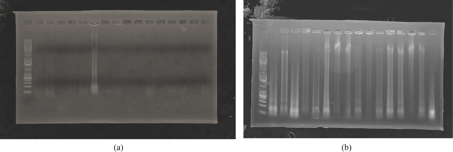
Clear differences in the DNA electrophoresis results from the wood and leafs of R. curtisii show the substantial challenges experienced in DNA extraction from woody tissues. Although a modified CTAB protocol was applied to address these challenges, the visual outcomes of DNA extraction from wood often show weak or undetectable bands, indicating technical difficulties that necessitate further exploration and refinement of extraction protocol. The absence of distinct DNA bands in wood samples does not necessarily show extraction failure. The band absence may reflect extremely low DNA concentrations, potentially due to the dense and lignified structure of wood, or the presence of inhibitors such as phenolic compounds and enzymes capable of interfering with downstream applications and hindering visualization. Therefore, conducting additional functional testing, including PCR, is essential to accurately assess the efficacy of the extraction methods used and confirm the presence of amplifiable DNA. During instances when DNA is not observable on electrophoresis gels, wood tissues remain a highly valuable DNA source for a variety of practical applications. Wood tissues are particularly relevant in species conservation where genetic information is crucial for aiding breeding programs and habitat restoration efforts. These are essential in the legal identification of illegally harvested log and processed wood forms, in order to support the enforcement of environmental regulations. Although the visual quality of DNA extracted from wood may currently fall short of ideal expectations, the established protocol provides a useful foundation for expanding molecular studies to include tissues that have previously posed significant genetic analysis challenges.
The need for modified DNA extraction procedures in wood tissues arises from the high concentrations of secondary metabolites, such as polyphenols and polysaccharides. The metabolites have a tendency to co-precipitate with DNA, leading to significant challenges in downstream molecular applications (Dumolin-Lapègue et al., 1999; Porebski et al., 1997) such as PCR and sequencing. Specifically, these compounds form complexes with nucleic acids, reducing the yield and purity of the DNA extracted while interfering with polymerase activity during PCR (Sharma et al., 2018). To mitigate the challenges, the optimized protocol incorporated the use of PVP and β-mercaptoethanol. PVP is known for the effective chelating properties, particularly targeting and binding polyphenols, thereby minimizing the inhibitory effects on DNA extraction. Similarly, β-mercaptoethanol plays a crucial role in breaking disulfide bonds in proteins, which can further complicate DNA extraction when not addressed (John, 1992). The addition of PVP and β-mercaptoethanol is critical for achieving successful wood DNA extraction due to the roles in neutralizing polyphenols and disrupting disulfide bonds in proteins, which are more abundant in wood than leafs (Rathore et al., 2025). Secondary metabolites, such as tannins and lignins, accumulate in older, lignified tissues of wood samples. This accumulation increases the complexity of the extraction process and significantly heightens the risk of PCR inhibition (Asif and Cannon, 2005), necessitating higher concentrations of PVP than the level primarily used for leaf samples. Therefore, the optimization of PVP concentration is essential for facilitating effective cell lysis and ensuring the purity of the extracted DNA.
This study showed the necessity of double TissueLyser processing times for wood tissues compared to leaf tissues due to the complex and rigid architecture of wood cell walls, which are composed of thick lignin and cellulose layers that resist mechanical disruption (Kurt et al., 2020). Leaf cells can be lysed relatively quickly due to the softer parenchyma structure, while wood tissues demand prolonged mechanical grinding periods, often requiring several minutes of rigorous bead beating to effectively break down the recalcitrant cell walls and facilitate the release of nucleic acids (Healey et al., 2014). The extended bead-beating duration improves DNA yield, which leads to higher concentrations of nucleic acids, but may increase fragmentation, necessitating careful optimization (Bürgmann et al., 2001).
Another critical consideration is the degradation of DNA in wood due to environmental exposure and the natural aging process, which further complicates extraction (Bürgmann et al., 2001; Rachmayanti et al., 2009). Compared to fresh leaves, wood samples may experience oxidative damage over time, leading to highly fragmented DNA, which can be hardly detected through traditional electrophoresis methods but still amplifiable in shorter target regions (Finkeldey et al., 2010; Jiao et al., 2020). This shows the importance of targeting smaller amplicons (e.g., chloroplast trnL-trnF) when working with wood-derived DNA (Schroeder et al., 2016). Given the ecological and economic importance of Dipterocarps, refining these methods is essential for advancing genetic study and forensic applications in tropical forestry, contributing to the conservation and sustainable management of the valuable forest ecosystems.
The effectiveness of the modified CTAB extraction protocol in this study was validated through PCR amplification of the chloroplast trnL-trnF marker from both wood and leaf-derived DNA of R. curtisii. According to Fig. 2(a) and (b), all samples had successful amplification, with PCR products showing distinct, single-copy bands for both tissue types. These results confirm that the optimized extraction method produced high-quality DNA suitable for amplification, despite the challenging nature of woody tissues. The clear banding patterns observed show that the modified protocol successfully overcame common obstacles associated with plant DNA extraction, particularly with recalcitrant wood samples.
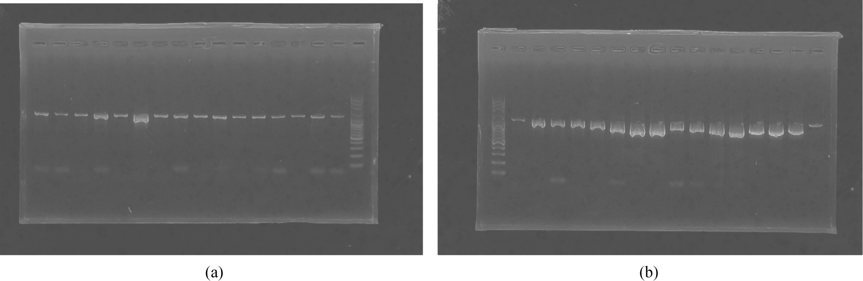
The visualization of DNA bands from wood tissue extracts was inconsistent in this study, and often faint or undetectable during agarose gel electrophoresis. However, the successful amplification through PCR showed the higher sensitivity of PCR compared to gel-based detection methods. This shows that the assessment of DNA quality for further molecular applications cannot depend solely on visualization results, suggesting a need for the inclusion of functional tests, such as PCR. A similar pattern was observed in the previous study on Rubroshorea leprosula (syn. Shorea leprosula), where the genomic DNA extracted from the wood did not produce visible results on electrophoresis gels. Effective amplification was carried out using PCR methods, specifically targeting several regions of chloroplast DNA (cpDNA; Rachmat et al., 2024b).
Successful PCR amplification using cpDNA markers from both leaf and wood-derived DNA in this study shows that the modified CTAB extraction protocol is capable of producing functional DNA compatible with further molecular analysis. The presence of clear, single, and specific PCR bands from both types of tissues shows that the challenging woody tissue extraction can be addressed using the modified method. However, the visual quality of DNA extracted from wood tissues remains a concern, particularly in the context of other molecular applications that require large amounts of DNA. More refinement of the extraction protocol, such as enhanced purification steps or buffer composition adjustments, may improve DNA recovery and consistency. Practically, these results reinforce the potential use of woody tissues as a DNA source for molecular-based species identification, which is highly relevant in the context of wood forensics, timber tracking, and conservation initiatives including R. curtisii and the Dipterocarpaceae family more broadly. Therefore, this study offers a practical solution in contexts where leaf material is unavailable, expanding the accessibility of molecular investigations to include previously intractable tissue types.
The chloroplast trnL-trnF intergenic spacer region was selected for PCR validation due to the established reliability for plant molecular studies, particularly when working with degraded or low-quality DNA samples. This combines several advantageous characteristics, including relatively short length (~300–500 bp), high copy number per cell, and conserved primer-binding sites flanking variable regions (Sarma et al., 2025). The characteristics make the spacer region exceptionally suitable for challenging templates such as wood-derived DNA, where fragmentation and inhibitor presence are common issues (Murillo-Sánchez et al., 2021). Furthermore, the trnL-trnF region has been widely used in Dipterocarpaceae phylogenetic studies (Carneiro de Melo Moura et al., 2019; Gamage et al., 2006; Kajita et al., 1998; Yulita et al., 2025), ensuring primer compatibility with R. curtisii. The success reported across various tissue types and preservation conditions (Murillo-Sánchez et al., 2021) contributed to the use of the trnL-trnF region as an ideal marker for validating DNA extraction protocol targeted at challenging plant materials.
Successful amplification of DNA from wood tissues in this study provides crucial alternatives for genetic studies on newly identified dipterocarp species, particularly given the considerable logistical challenges associated with accessing the tree canopy. Traditional methods of leaf sampling from towering trees such as R. curtisii often require dangerous climbing practices or the use of expensive canopy access equipment (Suhaimi et al., 2023), while wood samples can be collected more safely from fallen branches or logged materials. This method corresponds with growing applications of wood DNA in forest monitoring and timber tracking (Ng et al., 2022), particularly important for conservation of threatened dipterocarps (Tsumura et al., 2011). The protocol’s effectiveness with suboptimal samples enables the utilization of herbarium wood samples (Suhaimi et al., 2023), potentially unlocking historical genetic data. As leaf tissue remains the preferred source for high- quality DNA extraction, this wood-adapted method significantly expands study possibilities for tropical forest genetics and forensics.
BLASTn analysis of the chloroplast trnL–trnF sequences confirmed the successful molecular validation of genomic DNA extracted from both leaf and wood tissues. All sequences had 100% query coverage and e-values of 0.0, showing high-quality DNA suitable for downstream genetic analyses. Samples from the Bukit Tiban, Bukit Dangas, and Sei Harapan populations showed 100% sequence identity with Shorea curtisii reference sequences, signaling concordance between field identification and molecular data (Table 2).
Sequences from the Natuna Island population had equally high similarity (99.57%–99.79%) to several closely related Rubroshorea species rather than exclusively matching S. curtisii (Table 2). This pattern may reflect the conserved nature of cpDNA markers and the close evolutionary relationships within Dipterocarpaceae, where interspecific divergence in non-coding plastid regions is often low (Gamage et al., 2006; Heckenhauer et al., 2017; Hu et al., 2019). The limited variation observed among closely related taxa shows the restricted discriminatory power of the trnL–trnF region for species-level delimitation in this group.
In Dipterocarpaceae, several molecular barcoding and phylogenetic studies have reported limitations. For example, a comprehensive DNA barcoding assessment of lowland dipterocarps in Sumatra showed that the commonly used chloroplast markers (matK, rbcL, and trnL–F) were insufficient to resolve phylogenetic relationships within the Rubroshorea clade, despite matK possessing the highest discriminatory power among the tested loci. These results suggest that plastid barcodes alone have limited resolution for closely related species and additional loci or integrative methods are required for reliable species delimitation (Carneiro de Melo Moura et al., 2019).
Consistent BLAST results obtained from both leaf and wood-derived samples in each population show the robustness of the optimized CTAB protocol, particularly for woody tissues that are typically challenging due to high concentrations of secondary metabolites and polysaccharides. Although the trnL–trnF marker is not intended for definitive species identification, it provides reliable validation of DNA integrity and amplification success.
The sequence not only matches the type available in Genebank, but the clean and highly readable sequence obtained from the optimized DNA extraction protocol used in this study also clearly identifies the presence of single-nucleotide polymorphisms (SNPs), as shown in Table 3. The consistent nucleotide patterns observed in the wood-derived samples, particularly the patterns from Natuna (SCT_NTN_W1 and SCT_NTN_W2) and Batam (Bukit Tiban, Bukit Dangas and Sei Harapan), show that the extracted DNA retained sufficient integrity for downstream molecular analyses. Furthermore, the identical sequence patterns observed between wood and leaf samples from the same population suggest that the DNA obtained from wood tissues accurately represents the genetic information of the sampled individuals. These results present a consistent sequence pattern within populations but identify potential genetic differentiation between different population origin. Collectively, the results support the applicability of the modified CTAB method for both leaf and wood materials and show the potential for expanding genetic analyses in tropical tree biology, wood science, and forest ecology.
4. CONCLUSIONS
In conclusion, the modified CTAB extraction protocol developed for R. curtisii in this study had excellent performance, as evidenced by consistent PCR amplification success and the production of distinct single bands. In addition to successful amplification, the resulting sequences presented clear and readable electropherograms. Subsequent BLASTn validation confirmed the high quality of the obtained sequences, with 100% query coverage and strong similarity to reference sequences within Dipterocarpaceae. Sequence correlation further showed several polymorphic sites, including both SNPs and insertion–deletion events (indels), which distinguished two chloroplast haplotypes corresponding to the island of origin. The results showed that the modified CTAB protocol developed successfully produced DNA of sufficient quality for PCR amplification and downstream genetic analyses. This optimized method proves particularly valuable for extracting DNA from both wood and leaf tissues of ecologically important dipterocarp species. The protocol offers several key advantages, including (1) reliable DNA yield from challenging woody samples, (2) cost-effectiveness compared to commercial kits, and (3) broad applicability to other high-canopy dipterocarp species with similar sampling constraints. These characteristics make the protocol particularly suitable for establishing a DNA database and for forensic identification applications. The protocol can facilitate further genetic studies of R. curtisii, such as genetic profiling, phylogenetic analysis, and conservation studies, contributing to advancements in plant molecular investigations, where conventional leaf sampling methods are often impractical.