Journal of the Korean Wood Science and Technology
The Korean Society of Wood Science & Technology
Original Article

Fractionation of Essential Oils Extracted from Litsea angulata (Lauraceae) Using Two Different Methods and Their Impact on Lactic Acid Bacteria

Agmi Sinta PUTRI1,2, Irawan Wijaya KUSUMA1,2, Najmia Afifah ZULFA1, Rita DIANA3, Sarifah NURJANAH4, Kosei YAMAUCHI5, Harlinda KUSPRADINI1,2,https://orcid.org/0000-0001-5716-5955
1Laboratory of Forest Product Chemistry and Renewable Energy, Faculty of Forestry and Tropical Environment, Mulawarman University, Samarinda 75123, Indonesia
2Research Center of Medicine and Cosmetics from Tropical Rainforest (PUI-PT OKTAL), Mulawarman University, Samarinda 75119, Indonesia
3Laboratory of Ecology and Tropical Forests Biodiversity Conservation, Faculty of Forestry and Tropical Environment, Mulawarman University, Samarinda 75123, Indonesia
4Department of Agriculture Engineering and Biosystem, Padjadjaran University, Bandung 45363, Indonesia
5United Graduate School of Agriculture Sciences, Gifu University, Gifu 1121, Japan
Corresponding author: Harlinda KUSPRADINI (e-mail: hkuspradini@fahutan.unmul.ac.id)

Copyright 2025 The Korean Society of Wood Science & Technology. This is an Open-Access article distributed under the terms of the Creative Commons Attribution Non-Commercial License (http://creativecommons.org/licenses/by-nc/4.0/) which permits unrestricted non-commercial use, distribution, and reproduction in any medium, provided the original work is properly cited.

Received: May 20, 2025; Revised: Jun 16, 2025; Accepted: Jul 20, 2025

Published Online: Sep 25, 2025

ABSTRACT

This study was conducted to analyze the physicochemical characteristics of leaf essential oil fractions from Litsea angulata and evaluate their antibacterial activity against lactic acid bacteria. Essential oils were extracted and fractionated using two methods: simple fractional (SF) and spinning band (SB) distillation. Four bacterial strains were tested: Streptococcus mutans, Streptococcus sobrinus, Staphylococcus aureus, and Lactobacillus casei. The highest yields were obtained from fractions SF1 (0.83%, w/w) and SB6 (0.53%, w/w), which showed distinct color variations. Gas chromatography-mass spectrometry analysis revealed that monoterpenes, particularly α-phellandrene (> 40%), were the dominant components, followed by sesquiterpenes and other compounds. Fractions SF1 and SB6, which are rich in monoterpenes, exhibited the highest antibacterial activity, surpassing that of the positive control against S. mutans, S. sobrinus, and S. aureus. In contrast, SF2 (rich in sesquiterpenes, 33.36%) was more active against L. casei. These results suggest that L. angulata is a promising natural source of α-phellandrene and shows potential for development as an antibacterial agent, particularly for oral and dental health applications.

Keywords: fraction oil; simple fractional distillation; spinning band distillation; antibacterial activity

1. INTRODUCTION

Litsea, one of the largest genera in the Lauraceae family, is an important group of vegetation is widely distributed across tropical and subtropical forests (Jose et al., 2015; Tanaka et al., 2009; Wong et al., 2014). Species in this genus exhibit characteristic aromatic profiles and have long been used in traditional medicine to treat various conditions such as headaches, inflammation, intoxication, rheumatism, diarrhea, dysentery, spasms, wounds, fever, infections, bleeding, colic, vomiting, and other ailments (Chang and Chu, 2011; del Carmen Jiménez-Pérez et al., 2011; Hosamath, 2011; Rath, 2005; Yusuf et al., 2009).

Litsea species are also rich in bioactive compounds, including flavonoids (Lee et al., 2005; Wang et al., 2010), butanolides (Cheng et al., 2001, 2010), lignans (Pan et al., 2010), sesquiterpenes, fatty acids (Agrawal et al., 2011; Zhang et al., 2003), 1,3-diarylpropan-2-ol (Zhao et al., 2003), coumarins, syringaldehyde, and essential oils (Amer and Mehlhorn, 2006; Chowdhury et al., 2008; Guzmán-Gutiérrez et al., 2012). Essential oils from Litseaacutivena and Litseacubeba have demonstrated notable antibacterial and antifungal activity (Ho et al., 2011; Luo et al., 2004; Su and Ho, 2016). Various Litsea species exhibit antibacterial (Li et al., 2014; Wong et al., 2014), antifungal (Cruz et al., 2014; Pambudi et al., 2018), antioxidant (Arfan et al., 2008; Wulandari et al., 2018), cytotoxic (Feng et al., 2009), and anti-inflammatory properties (Gogoi et al., 2018).

Wang and Liu (2010) reported that various parts of L. cubeba, including the fruits, leaves, roots, buds, stems, and flowers, can produce essential oils rich in citral (B), β-phellandrene, and β-terpinene. The leaf oil of this species shows strong bactericidal effects against Escherichia coli (Nguyen et al., 2017) and antimicrobial activity against cariogenic bacteria such as Streptococcus mutans, Streptococcus sobrinus, and Streptococcus sanguinis (Yang et al., 2013). Similarly, the leaf oil of Litseapetiolata inhibits several pathogenic bacteria, including Bacillus cereus, Bacillus subtilis, Staphylococcus aureus, Staphylococcus epidermidis, Streptococcus agalactiae, Vibrio cholerae, Vibrio parahaemolyticus, and Salmonella typhimurium (Thongthip et al., 2017). Ho et al. (2011) also found that the leaf and root oils of L. acutivena and the leaf oil of L. coreana possess strong antimicrobial and anti-wood rot activities. In addition, the leaf and bark extracts of Litsea glutinosa exhibited antibacterial activity against S. aureus, S. epidermidis, E. coli, Enterobacter intermedium, and Salmonella spp. (Haque et al., 2014).

Litsea angulata is native to Borneo, Sumatra, Java, and New Guinea and is commonly found in the forests of East Kalimantan. In South Korea, it is traditionally used to treat boils and inflammation (Ariyani and Mustikasari, 2010). Previous studies reported that seed extracts of L. angulata contain alkaloids and tannins and have potential as natural spermicides (Akmal et al., 2016). Saputri and Susiani (2018) found that the fruits and seeds of L. angulata exert antioxidant activity. Kuspradini et al. (2019) demonstrated that the bark, branches, and leaves of this species are rich in natural antioxidants and possess antibacterial properties. Crude essential oils extracted from the leaves of L. angulata also show antimicrobial activity against S. mutans, S. sobrinus, S. aureus, and Candida albicans (Kuspradini et al., 2020). In contrast to previous studies using crude extracts, we employed two different distillation techniques, simple fractional (SF) and spinning band (SB) distillation, to separate and isolate specific fractions of the essential oil. This approach enables a more precise understanding of the bioactive constituents that contribute to antibacterial efficacy.

Despite advancements in medicine, microbial infections, including those caused by bacteria, viruses, parasites, and fungi, remain important public health issues in many countries (Jawetz et al., 2005). Among these, dental caries is one of the most prevalent infectious diseases worldwide (Chaiya et al., 2013; Forssten et al., 2010), greatly contributing to the burden of oral health problems (Widayati, 2014). Dental caries typically develop when food residues adhere to teeth, promoting calcification, decay, and eventual tooth damage (Sinaga, 2013). The main causative agents include lactic acid bacteria, particularly S. mutans and Lactobacillus spp.

Although lactic acid bacteria support digestive health, they can contribute to tooth decay by fermenting dietary carbohydrates and producing acids that erode enamel. These bacteria become active shortly after consumption, forming biofilms and plaques. To prevent these actions, topical antimicrobial agents such as mouthwashes and toothpaste are used. Synthetic antibiotics, including penicillin, vancomycin, and chlorhexidine, can lead to side effects, such as antibiotic resistance and dental discoloration (Van Winkelhoff et al., 2000).

In response to these concerns, natural antibacterial compounds are being increasingly evaluated as safer alternatives. Plant-derived essential oils are particularly attractive because of their low risk of adverse effects (Indrawati et al., 2017).

Studies of the antibacterial potential of L. angulata essential oil, particularly its individual chemical fractions, are limited. Therefore, this study was conducted to (1) fractionate L. angulata essential oil using both simple SF and SB distillation, (2) identify the chemical constituents of each fraction, and (3) evaluate and compare their antibacterial activities against oral pathogens. This approach allows for detailed analysis of how the fractionation method influences chemical composition and bioactivity.

2. MATERIALS and METHODS

2.1. Plant and chemical materials

Fresh leaves of L. angulata were collected from the Educational Forest (KHDTK Lempake), Faculty of Forestry and Tropical Environment, Mulawarman University, Samarinda, East Kalimantan, Indonesia (00°26’55.1”S, 117°12’36.2”E). The experiments were conducted in 2018 at the Forest Product Chemistry and Renewable Energy Laboratory, Faculty of Forestry and Tropical Environment, Mulawarman University. A specimen of the plant was deposited as a voucher in the Dendrology Laboratory of Mulawarman University.

Analytical-grade chemicals such as nutrient broth, glucose, and anhydrous sodium sulfate were purchased from Merck (Darmstadt, Germany). All other reagents used were commercial standards.

2.2. Simple fractional distillation

Essential oils were extracted via water and steam distillation from 4,200 g of fresh L. angulata leaves. The leaves were air-dried for 1 day. Fractionation was performed at distillation time intervals of 0–60 min (SF1), 60–120 min (SF2), and 120–180 min (SF3) as described by Zheljazkov et al. (2014) with slight modifications.

At the end of each interval, the oil fractions were separated from the water, and any remaining moisture was removed using anhydrous sodium sulfate. The yield of each fraction was calculated as the percentage of the oil volume relative to the dry weight of the plant material. The essential oils were stored in sealed vials in the dark until further analyses.

2.3. Spinning band distillation

The essential oil from L. angulata leaves was extracted using steam distillation method (Kuspradini et al., 2016; Putri et al., 2018). The samples were air-dried at room temperature for one day. The essential oils were prepared for further analyses. The leaf oil was fractionated using the SB distillation method with a B/R Instrument-Spinning Band Distillation System Model 36-100 (Amrullah et al., 2017). Approximately 50 mL (43.88 g) of essential oil was added to a boiling flask connected to a fractionating column 90 cm in length and controlled by a heat mantle and condenser system (35°C). This process was maintained under vacuum conditions using a vacuum pump with the following settings: pressure 10 mmHg, initial heat 24%, equilibration time 15 min, maximum pot temperature 300°C, and heat rate 16%. The fraction was separated according to the boiling points (60°C–300°C). The volume of various fractions is presented as a percentage of the yield (% w/w; Torres et al., 2012).

2.4. Gass chromatography-mass spectrometry analysis

The chemical composition of each oil fraction was analyzed using gas chromatography-mass spectrometry (GC-MS; QP-2010, Shimadzu, Kyoto, Japan) equipped with an RTX-5MS capillary column (30 m × 0.25 mm × 0.25 μm). The column conditions were as follows Iordache et al. (2011): injector temperature of 250°C, detector temperature of 280°C, inlet pressure of 93.5 kPa, split ratio of 200:1, column flow rate of 1.44 mL/min, linear velocity of 49.3 cm/s, and helium as a carrier gas.

Compounds were identified by comparing their mass spectra with the NIST database and by calculating retention indices (RI) using C8–C20 alkane standards as described by van Den Dool and Dec. Kratz (1963). Each analysis was performed in duplicate.

2.5. Antibacterial activity measurement

The essential oil fractions were screened for their antibacterial activity using the agar well diffusion method, according to the method described by Kuspradini et al. (2018). The bacterial strains used were S. mutans (KCCM 40105), S. sobrinus (KCCM 11898), S. aureus, and Lactobacillus casei. Briefly, the plates were inoculated with the bacterial suspension under aseptic conditions; the 7-mm diameter wells were filled with 20 μL of the samples. The agar plates were incubated at 37°C for 18–24 h (Su et al., 2015). The turbidity of the bacterial suspension was measured at 600 nm (Jahangirian et al., 2013). Chloramphenicol [K(+)1] and chlorhexidine [K(+)2; concentrations of 0.001% or 0.01 mg] were used as standard antibiotics for the positive control, and 40% ethanol was used as the negative control. The concentrations of essential oil used in this test were 100%, 50%, and 25% measured in 20 μL of oil and converted to mg (mg/20 μL) as shown in Table 1. The study was performed in triplicate. After incubation, the diameter of inhibition of bacterial growth was observed and measured in millimeters using a ruler. The data are presented as the average of three replicate experiments.

Table 1. Concentrations of essential oil fractions
Samples Concentrations Samples Concentrations
% mg/20 μL % mg/20 μL
Crude oil 100 19.1
Simple fractional distillation (SF) Spinning band distillation (SB)
SF1 100 17.4 SB5 100 17.9
50 8.7 50 8.95
25 4.35 25 4.475
SF2 100 17.5 SB6 100 21.7
50 8.75 50 10.85
25 4.375 25 5.425
SF3 100 16.5 SB7 100 21.7
50 8.25 50 10.85
25 4.125 25 5.425
Download Excel Table

3. RESULTS and DISCUSSION

3.1. Fractionation by simple fractional distillation and spinning band distillation

Fractional distillation separates essential oils into several fractions. During this process, some variables can be used as treatments to increase the quality of the oil (Amrullah et al., 2017). The aim of this study was to separate L. angulata leaf essential oils into distinct fractions to enhance the oil quality. Two methods were employed: SF distillation (based on distillation time) and SB distillation (based on boiling point ranges).

SF distillation was performed using the water and steam distillation method for different distillation times: 0–60 min (SF1), 60–120 min (SF2), and 120–180 min (SF3). The air-dried material (4,200 g) prepared one day prior as described in section 2.2 was used as the raw material. The yield was measured based on the distribution of the fraction weight with the raw material weight (oven dry/OD), which was multiplied by 100 to obtain the percentage yield.

SB distillation was performed based on the boiling points of the main components of the pure oil. L. angulata leaves were distilled to extract essential oil, which was then fractionated using an SB tool. The essential oil (± 50 mL, 43.88 g, 1.05%) was subjected to the oil distillation process three times, with each process lasting for ± 3–4 h. The yield of each fraction was measured based on the relationship between the fraction weight and essential oil weight (w/w), which was then converted to the initial oil yield weight (Torres et al., 2012).

Fractional distillation of L. angulata leaf essential oil with SF and SB distillation yielded three oil fractions. The fractions produced by each method had various yields (%) and colors, as presented in Table 2.

Table 2. Yield and color of Litsea angulata leaf oil fractionated by simple fractional distillation (SF) and spinning band distillation (SB)
Samples Treatments Yields (%) Color
SF Distillation time (min)
SF1 0–60 0.83 Clear
SF2 60–120 0.25 Yellowish
SF3 120–180 0.07 Greenish
SB Temperature range (°C)
SB1 60–67 - -
SB2 67–78 - -
SB3 78–82 - -
SB4 82–160 - -
SB5 160–170 0.11 Yellowish
SB6 170–210 0.53 Clear
SB7 210–220 0.08 Orange
SB8 220–300 - Dark orange (residue)
Download Excel Table

The results of the SF distillation showed that the highest yield was obtained in fraction SF1 (0.83%), whereas the lowest yield was obtained in fraction SF3 (0.07%; Table 2). This approach was used to isolate essential oil into fractions in a simple manner. This method is effective for obtaining L. angulata oil quickly, as the highest yield was obtained from the first distillation, followed by a second fraction within 60–120 min; thus, the entire process was completed in 0–120 min. Water and steam distillation is a simple method for obtaining essential oils in a short distillation time, high yield, and high quality. This distillation system is widely used because of the low potential for oil decomposition (ester hydrolysis, polymerization, resinification; Qin et al., 2014; Stratakos and Koidis, 2015; Turek and Stinzing, 2013).

The fractionation results from SB distillation in Table 2 show that the oil did not separate between temperatures of 60°C–160°C but did separate in the temperature range of 160°C–220°C. The SB6 fraction showed the highest yield (0.53%), followed by SB5 (0.11%), and SB7 (0.08%). During this process, residual non-distilled materials (SB8) remained in a paste form, even at the highest temperature. According to Savakar et al. (2017), SB distillation involves the use of a metal column to isolate samples with a broad range of boiling points. The temperature determined during the distillation process is the component distillation point; thus, the distillation temperature range matched the boiling point of the constituent material components. Using higher temperatures leads to decomposition and resinification of the produced distillate (Amrullah et al., 2017; Mangun et al., 2005).

Visual observation of the oil fraction of L. angulata showed that the color of oil produced using each method varied. The oil first obtained by SB fractional distillation exhibited a different color than that observed in the test results, as the fractions were oxidized quickly, resulting in a color change. Fraction SB7 was orange in color, possibly because of its high boiling point (210°C–220°C); therefore, the SB method is not recommended for samples with high boiling points to ensure good color quality. The color variation of the fractionated oil may reflect differences in the chemical composition, particularly the content of volatile compounds such as monoterpenes and sesquiterpenes (Amrullah et al., 2017). Supriono and Susanti (2014) observed that darker patchouli oil tended to have a lower alcohol content, in contrast, in our study, fraction SB6—despite its darker color—contained a high proportion of α-phellandrene and showed strong antibacterial activity. Essential oils frequently appear clear and fluid; however, some oils, such as Orris, are solid, whereas others, such as guaiac wood, are semi-solid at room temperature. Most essential oils are either colorless or pale yellow; however, some oils, such as blue chamomile and European valerian, exhibit deeper colors, with the latter being green (Tisserand and Young, 2013). Factors such as the drying of raw materials and extraction procedure may influence the essential oil color (Ahmad et al., 2014; Rahimmalek and Goli, 2013).

3.2. Chemical compositions of oil fractions determined using gas chromatography-mass spectrometry analysis

The constituent chemical components of each oil fraction of L. angulata obtained from both fractional distillation methods were analyzed using GC-MS. GC-MS analysis is a rapid and accurate method for separating complex mixtures, analyzing mixtures in small amounts, and obtaining data on the structure and identity of organic compounds (Agusta, 2000). Because essential oils contain volatile compounds, they can be analyzed using GC-MS. The Kovats retention index value (RI) of the identified compounds was compared with the reference RI in NIST to confirm the results of compound identification, and the RI was calculated by comparison with the alkane standard (C8–C20).

The constituent compounds identified in each fraction of L. angulata leaf essential oil using SF and SB distillation are shown in Tables 3 and 4. Based on the GC-MS results, each oil fraction of L. angulata contained different main constituent compounds, although the dominant compounds were the same between SF and SB distillation, and some compounds were present in samples obtained using both methods.

Table 3. Chemical constituents of Litsea angulata leaf essential oil fractions by simple fractional distillation
Chemical components Group Percentage (%) Retention indices (RI)
SF1 SF2 SF3 SF1 SF2 SF3 Ref1)
β-Pinene MH 2.59 ± 0.26 1.17 ± 0.25 - 948 950 - 952
β-Myrcene MH 1.03 ± 0.04 - - 958 - - 962
α-Phellandrene MH 47.97 ± 4.82 15.22 ± 1.36 2.12 ± 0.11 999 955 1,000 1,001
β-Cymene MH 6.13 ± 0.43 7.26 ± 0.53 3.87 ± 0.31 1019 1,019 1,060 1,022
β-Phellandrene MH - 2.75 ± 0.33 - - 1,026 - 1,032
(.+-.)-Camphor MO - 1.17 ± 0.35 - - 1,145 - 1,144
A-phellandrene epoxide MO - 1.59 ± 0.03 - - 1,198 - -
α-Terpinolene MH 2.46 ± 0.26 1.28 ± 0.12 - 1,082 1,082 - 1,089
Limonene dioxide 1 MO 5.36 ± 0.79 6.09 ± 0.81 - 1,200 1,200 - -
Acetic acid 1,3,3-trimethyl-2-oxa-bicyclo[2.2.2]oct-6-yl ester O 10.25 ± 2.86 12.56 ± 1.51 3.23 ± 0.19 1,311 1,313 1,314 1,431
α-Copaene SH - 2.79 ± 0.08 8.98 ± 0.11 - 1374 1,375 1,376
δ-Elemene SH - - 1.58 ± 0.12 - - 1,331 1,343
cis-p-Mentha-1(7),8-dien-2-ol MO 4.35 ± 1.34 4.24 ± 0.63 - 1,386 1,387 - -
2-Cyclohexen-1-one, 3-(hydroxymethyl)-6-(1-methylethyl)- MO 3.90 ± 0.78 - - 1,464 - - -
trans-Caryophyllene SH 3.96 ± 0.49 11.92 ± 0.23 31.43 ± 0.24 1,563 1,570 1,598 -
cis-p-Mentha-1(7),8-dien-2-ol MO - 1.23 ± 0.07 - - 1,729 - -
Aromadendrene VI SH - - 0.93 ± 0.01 - - 1,738
α-Humulene SH - 1.52 ± 0.31 4.96 ± 0.09 - 1,780 1,772 -
(+)-Cycloisosativene SH - - 3.42 ± 0.14 - - 1,651
α-Gurjunene SH - 0.99 ± 0.35 5.05 ± 0.06 - 1,580 1,573 -
α-selinene SH - - 2.61 ± 0.08 - - 1,512 1,517
δ-Cadinene SH - - 4.30 ± 0.12 - - 1,521 1,523
Palustrol SO - 1.33 ± 0.03 2.14 ± 0.03 - 1,578 1,580 1,570
(-)-Caryophyllene oxide SO - 10.07 ± 0.25 12.74 ± 0.04 - 1,590 1,592 1,581
Epiglobulol SO - - 0.93 ± 0.06 - - 1,681 -
(-)-Caryophyllene oxide SO - 1.16 ± 0.01 1.65 ± 0.03 - 1,951 1,970 1,581
(+)-Aromadendrene SO - 2.11 ± 0.13 - - 1,966 - -
Epiglobulol SO - - 4.21 ± 0.08 - - 1,962 -
Torreyol SO - 1.47 ± 0.02 2.68 ± 0.26 - 1,917 1,914 -
MH 60.17 27.68 5.99
MO 13.61 14.32 0
O 10.25 12.56 3.23
SH 3.96 17.22 63.24
SO 0 16.14 24.34

Data from NIST (2019).

SF: simple fractional distillation, MH: monoterpene hydrocarbons, MO: oxyterpene oxygenation, O: others; SH: sesquiterpene hydrocarbons, SO: oxygenated sesquiterpenes.

Download Excel Table
Table 4. Chemical constituents of Litsea angulata leaf essential oil fractions by spinning band distillation (SB)
Chemical components Group Percentage (%) Retention indices (RI)
SB5 SB6 SB7 SB5 SB6 SB7 Ref1)
β-Pinene MH 4.36 ± 0.98 0.38 ± 0.02 - 955 954 - 952
β-Myrcene MH 0.66 ± 0.13 - - 964 - - 962
α-Phellandrene MH 29.28 ± 3.38 21.44 ± 2.26 2.73 ± 0.18 1,005 1,006 1,002 1,001
δ-3-Carene MH 0.82 ± 0.18 - - 1,010 - - 1,010
α-Terpinene MH 1.21 ± 0.25 1.40 ± 0.25 - 1,014 1,015 - 1,014
p-Cymene MH 7.53 ± 1.29 7.87 ± 0.89 1.94 ± 0.06 1,022 1,024 1,021 1,022
(-)-Limonene MH - 3.43 ± 0.25 - - 1,028 - 1,027
trans-β-Ocimene MH - 0.43 ± 0.11 - - 1,040 - 1,047
γ-Terpinene MH - 0.90 ± 0.13 - - 1,055 - 1,058
α-Terpinolene MH - 2.74 ± 0.35 1.13 ± 0.06 - 1,086 1,085 1,063
3-Octen-2-one. 7-methyl- O - 0.39 ± 0.06 - - 1,096 - -
trans-caran. 4.5-epoxi- MO - 1.04 ± 1.07 - - 1,131 - -
p-menth-2-en-1-ol MO - - 0.63 ± 0.02 - - 1,138 1,137
1.3-Dimethyl-1-cyclohexene O - - 1.35 ± 0.04 - - 1,171
Carvenone MO - 0.61 ± 0.00 - - 1,171 -
4-Terpineol MO - - 1.02 ± 0.01 - - 1,177 1,177
trans-2.3-Epoxicaran MO - 0.71 ± 0.03 - - 1,178 -
trans-p-Mentha-1(7).8-dien-2-ol MO 0.52 ± 0.01 - - 1,183 - 1,185
p-Cymen-8-ol MO - 0.97 ± 0.06 1.65 ± 0.04 - 1,183 1,182 1,183
α-Terpineol MO - - 0.86 ± 0.04 - - 1,189 1,187
Decanal MO - - 1.02 ± 0.00 - - 1,194 1,208
A-Phellandrene Epoxide MO 2.03 ± 0.22 1.95 ± 0.25 4.83 ± 0.07 1,200 1,200 1,200 -
1.8-Cineole MO 1.35 ± 0.23 1.17 ± 0.23 - 1,200 1,200 - -
cis-Sabinol MO - - 1.22 ± 0.03 - - 1,200 1,170
Limonene dioxide 4 MO 6.47 ± 0.71 7.50 ± 1.38 - 1,200 1,200 - -
Carvotanacetone MO - - 1.57 ± 0.04 - - 1,200 1,230
Piperitone MO - - 0.49 ± 0.00 - - 1,200 1,227
Ascaridole MO 0.74 ± 0.26 - - 1,200 - - 1,237
Thujyl alcohol isomer MO - 0.52 ± 0.30 -
-
- 1,200 - -
Bicyclo[3.3.1]hept-3-en. 4.6.6-trimethyl-2-vinyloxy- MO 0.53 ± 0.01 - - 1,245 - - -
1-ethoxymethyl-4-methylene-cyclohexane MO - - 0.83 ± 0.05 - - 1,204 -
Phenol. 3-methyl-5-(1-methylethyl)-.methylcarbamate O - - 1.18 ± 0.01 - - 1,251 -
cis-Limonene oxide MO - 1.29 ± 0.47 - - 1,235 -
Thujyl alcohol MO - - 0.39 ± 0.01 - - 1,301 -
Acetic acid 1.3.3-trimethyl-2-oxa-bicyclo[2.2.2]oct-6-yl ester O - 0.90 ± 0.11 1.21 ± 0.08 - 1,278 1,309 1,341
1.3.3-trimethyl-2-oxabicyclo [2.2.2]octan-6-ol MO 7.65 ± 0.54 - 3.55 ± 0.53 1,320 - 1,318 -
Ascaridole MO 5.04 ± 0.20 8.64 ± 3.92 - 1,328 1,330 - 1,237
α-Copaene SH - - 26.13 ± 0.31 - - 1,382 1,379
(-)-β-Elemene SH - - 0.77 ± 0.04 - - 1,391 1,391
Limonene dioxide 1 MO - - 2.06 ± 0.00 - - 1,478 -
cis-Piperitone oxide MO 4.72 ± 0.17 4.63 ± 0.02 - 1,657 1,537 - -
trans-Caryophyllene SH - - 0.98 ± 0.05 - 1,598 -
trans-6-hydroxy-p-menth-1-en-3-one MO 1.84 ± 0.07 - - 1,700 - - -
α-Bergamotene SH - - 1.59 ± 0.01 - - 1,627 -
α-Guaiene SH - - 0.63 ± 0.01 - - 1,683 -
α-Humulene SH - - 1.65 ± 0.11 - - 1,753 -
Alloaromadendrene SH - - 1.04 ± 0.01 - - 1,703 -
α-Amorphene SH - - 2.53 ± 0.02 - - 1,648 -
2-isopropenyl-4a.8-dimethyl-1.2.3.4.4a.5.6.7-octahydro-naphthalene SH - - 0.79 ± 0.04 - - 1,572 -
β-Selinene SH - - 3.97 ± 0.06 - - 1,533 1,527
β-Bisabolene SH - - 12.47 ± 0.23 - - 1,502 1,502
Diepi-.alpha.-cedren I SH - - 1.03 ± 0.06 - - 1,513 -
γ-Muurolene SH - - 1.49 ± 0.03 - - 1,520 1,515
δ-Cadinene SH - - 9.79 ± 0.09 - - 1,527 1,527
Naphthalene. 1.2.3.4.4a.7-hexahydro-1.6-dimethyl-4-(1-methylethyl)- SH - - 0.80 ± 0.04 - - 1,538 1,539
α-Muurolene SH - - 0.44 ± 0.01 - - 1,543 1,540
Patchouli alcohol SO 1.58 ± 0.01 1.04 ± 0.06 - 1,768 1,768 -
trans-Pinocarveylacetate O - - 1.68 ± 0.10 - - 1,720 -
Solanone O 0.99 ± 0.92 - - 1,652 - - -
Limonene dioxide 3 MO 1.12 ± 0.38 - - 1,614 - - -
cis-Limonene oxide MO 1.66 ± 0.49 1.69 ± 0.54 - 1,644 1,645 - -
MH 43.84 38.58 5.80
MO 33.65 30.70 20.09
O 0.99 1.28 5.42
SH 0.00 0.00 66.56
SO 1.58 1.04 0.00

Data from NIST (2019).

MH: monoterpene hydrocarbons, O: others, MO: oxyterpene oxygenation, SH: sesquiterpene hydrocarbons, SO: oxygenated sesquiterpenes.

Download Excel Table

The chemical compounds identified in fractions SF1–SF3 are shown in Table 3. Based on the GC-MS data, the dominant compounds in fraction SF1 were monoterpenes, with main compounds including α-phellandrene (47.97%), acetic acid 1,3,3-trimethyl-2-oxa-bicyclo[2.2.2]oct-6-yl ester (10.25%), and β-cymene (6.13%). The two dominant chemical components in fraction SF2 were monoterpenes (42%) and sesquiterpenes (33.36%). The four main compounds were α-phellandrene (15.22%), acetic acid 1,3,3-trimethyl-2-oxa-bicyclo[2.2.2]oct-6-yl ester (12.56%), trans-caryophyllene (11.92%), and (-)-caryophyllene oxide (10.07%). Fraction SF3 was dominated by sesquiterpenes, as well as trans-caryophyllene (31.43%), (-)-caryophyllene oxide (12.74%), and α-copaene (8.98%).

The distillation time can also affect the quantity and quality of the essential oil. The duration of essential oil extraction significantly influences both the yield and proportion of chemical compounds present in the oil (Oliveira et al., 2017). Chatzopoulou and Katsiotis (1995) and Prins et al. (2006) evaluated the influence of extraction time on the chemical composition of essential oils and detected the concentration of oxygenated compounds at the beginning of the extraction process. The chemical components and amounts contained in the distillation results were diverse. Low-boiling point components such as α-pinene, camphene, β-pinene, and myrcene were present at higher concentrations at the beginning of the distillation but decreased over time, reaching a minimum at the end of distillation (Zheljazkov et al., 2014). Other compounds in the sesquiterpene groups in essential oils were found in greater amounts and required longer distillation times (up to 5 h) because the molecules were non-polar (Oliveira et al., 2017).

Table 4 presents the GC-MS data for the oil fraction obtained using SB distillation. The chemical compounds identified in fraction SB5 were dominated by hydrocarbon monoterpene (43.84%), oxygenated monoterpene (33.65%), oxygenated sesquiterpene (1.58%), and other compounds (0.99%). Three main compounds in fraction SB5 were α-phellandrene (29.28%), p-cymene (7.53%), and 1.3.3-trimethyl-2-oxabicyclo[2.2.2]octan-6-ol (7.65%). The main constituents of this fraction are classified as hydrocarbon monoterpene and oxygenated monoterpene compounds. GC-MS analysis showed that the main compounds in fraction SB6 consisted of three peak compound components included in hydrocarbon monoterpene family, namely α-phellandrene (21.44%), p-cymene (7.87%), and oxygenated monoterpene, specifically ascaridole (8.64%). Fraction SB7 was dominated by sesquiterpene group compounds (66.56%) followed by oxygenated monoterpenes (20.09%), other compounds (5.42%), and hydrocarbon monoterpenes (5.80%). Three main components of peak compounds in this fraction were α-copaene (26.13%), β-bisabolene (12.47%), and δ-cadinene (9.79%), which are all members of the sesquiterpene group.

Fractional distillation is used to separate crude oil into useful substances (or fractions) containing different hydrocarbons at different boiling points. A crude oil fraction with a higher boiling point has a larger number of carbon atoms, higher molecule weight, darker color, and lower viscosity compared to these factors in fractions with lower boiling points (Kumar and Tripathi, 2011). Maiwald and Schwantes (1991) isolated essential oils from Curcuma rhizomes based on their boiling points. Compounds in the essential oil contain monoterpenes with boiling points of 140°C–180°C, such as 1.8-cineol and α-phellandrene, and sesquiterpenes with boiling points of > 200°C, such as β-bisabolene. α-Phellandrene is a natural compound that can stimulate the immune response in leukemia model rats (Lin et al., 2014). Additionally, α-phellandrene exerts effects against human breast cancer and prostate tumor cells (Essien et al., 2012). Monoterpenes [such as eucalyptol, borneol, camphor, bornyl acetate, carvacrol, menthol, γ-terpinene, (+)-α-pinene, (-)-β-pinene, and p-cymene] are pivotal components of essential oil produced through liquid extraction and distillation with steam from edible plant species and medicinal plants (Crozier et al., 2006).

3.3. Antibacterial activity of Litsea angulata oil fractions

L. angulata crude essential oil and its fractions were evaluated for their antibacterial activity in vitro using the agar well diffusion method. This method is based on the antibacterial abilities of compounds, which influence the diameter of the inhibition zone around the test bacteria (Nurainy et al., 2008). In most cases, the Standard Deviation (SD) values were relatively small, indicating good reproducibility of the results. However, some treatments showed larger SD values, suggesting variability in the bacterial response or sample sensitivity. In this study, two types of antibiotics were used as positive controls: chloramphenicol and chlorhexidine. Chloramphenicol is an antimicrobial agent that can be used to treat cocci and basil gram-positive bacteria, as well as aerobic and anaerobic gram-negative gram bacteria (U.S. Department of Health and Human Services, 2000). Chlorhexidine is among the most common and widely used antiplaque and anti-gingival agents (Balagopal and Arjunkumar, 2013; Mathur et al., 2011). The inhibition activity of every L. angulata oil fraction at concentrations of 100%, 50%, and 25%, which were converted to mg/20 μL, against the four test bacteria was determined by measuring the clear zone. The results indicated that the oils exerted antibacterial activities. The inhibition diameters created by the L. angulata essential oil fractions against S. mutans, S. sobrinus, S. aureus, and L. casei compared to those created by the crude oil and synthetic standards are presented in Fig. 1. Each fraction obtained using the SF distillation method exhibited antibacterial activities against these four bacteria.

wood-53-5-501-g1
Fig. 1. Antibacterial properties of Litsea angulata leaf crude essential oil and fractions by simple fractional distillation (SF) against. (a) Streptococcus mutans, (b) Streptococcus sobrinus, (c) Staphylococcus aureus, (d) Lactobacillus casei. The inhibition assays were performed in triplicate, and the error bars represent the standard deviation (SD) of the measurements.
Download Original Figure

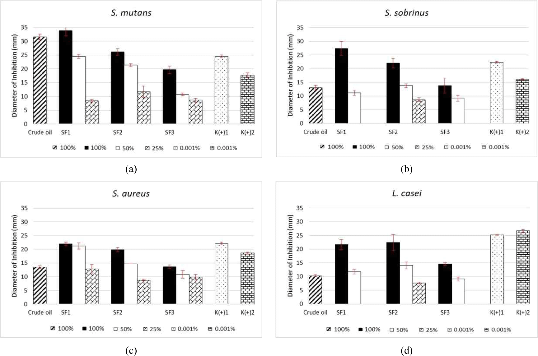
All fractions examined at the three concentrations highly inhibited S. mutans [Fig. 1(a)], with decreasing effectiveness as the fraction concentration was decreased (100% > 50% > 25%). Each pure fraction inhibited the growth of S. mutans, with the highest inhibition value observed for the SF1 fraction (33.89 mm), followed by SF2 (26.11 mm) and SF3 (19.67 mm). These results indicate that at a concentration of 50%, the samples inhibited bacteria compared to the same extent as both positive controls, with a value of 24.56 mm for the SF1 fraction. This inhibition value was equal to that of chloramphenicol. High inhibition was also observed for the 50% SF2 fraction (21.33 mm), whereas at a concentration of 25%, the inhibition ability of the three fractions did not surpass the activity of the standards. The high inhibition value of the SF1 fraction was likely related to the dominant compound in the fraction, α-phellandrene (47.97%), as well as other compounds in the monoterpene group. Işcan et al. (2012) reported that α-phellandrene exerts antimicrobial, antibacterial, and fungicidal activities. Subsequently, İşcan (2017) found that α-phellandrene of the hydrocarbon monoterpene group inhibited Enterobacter aerogenes, Serratia marcescens, E. coli, S. typhimurium, B. cereus, S. aureus, Listeria monocytogenes, and Candida spp. Ghasemi Pirbalouti and Gholipour (2016) and Buitrago et al. (2012) found that an essential oil with antibacterial activity contained > 30% α-phellandrene as the main compound. According to Zhang et al. (2017), the mycelia growth of P. cyclopium was significantly inhibited by α-phellandrene and nonanal by severely disrupting the integrity of the fungal cell membrane, resulting in leakage of cell constituents and potassium ions. Additionally, these substances trigger an increase in total lipid content, extracellular pH, and membrane permeability.

The largest inhibition value (27.33 mm) in the antibacterial test against S. sobrinus was observed for the SF1 fraction; however, inhibition drastically decreased at a concentration of 50%, with no inhibition at 25% (Fig. 1). Fraction SF2 showed the second-largest inhibition value, with an inhibition diameter of 22.00 mm, which was higher than that of the chlorhexidine positive control. Similar to SF1, 25% SF3 exhibited no activity against the tested bacteria. In addition to the dominant α-phellandrene, the SF1 fraction contained p-cymene in monoterpene group as a main compound. p-Cymene is the main antimicrobial compound found in thyme. Considerable evidence suggests that monoterpenes have antibacterial, antivirus, and antifungal activities. Furthermore, p-cymene inhibits tumor proliferation through antiangiogenic mechanisms, cytotoxicity against cancer cells, and anti-adhesion action (Kaluđerović et al., 2015; Li et al., 2016; Păunescu et al., 2015). Recent observations indicate that p-cymene decreases biofilm development by Burkholderia xenovorans by accumulating in bacterial membranes and altering the membrane structure (Agulló et al., 2017). Additional studies indicated that hydrocarbons and oxygenated monoterpenes possess a wide spectrum of antimicrobial activities against pathogenic microorganisms (Badawy et al., 2019; Garcia et al., 2008; Marei et al., 2019; Niksic et al., 2017).

Analysis of S. aureus, showed that the SF1 pure fraction was the most active inhibitor, with an inhibitor diameter of 22.00 mm; this fraction was also active at a concentration of 50% [21.22 mm; Fig. 1(c)]. This result indicates that this diluted fraction also has inhibition effects. For SF2, the inhibition zone was larger than that of chlorhexidine, which was 19.89 mm (pure fraction). All concentrations applied to S. aureus inhibited the bacteria by up to 25%. SF1 showed the second largest inhibition zone at a 50% concentration, followed by SF2 with an inhibition value of 14.67 mm, whereas the smallest was observed for SF3 (10.89 mm). At a concentration of 25%, SF1 achieved the largest inhibition value of 12.89 mm, whereas the lowest value was observed for SF2 (8.78 mm). These results indicate that SF1 had the strongest activity at a concentration of 25% (12.89–22.00 mm). In North Kalimantan, L. angulata plants are traditionally used to cure blains, a skin infection commonly caused by S. aureus (Ariyani and Mustikasari, 2010; McCaig et al., 2006). Kuspradini et al. (2019) showed that the leaves, bark, and branches of L. angulata inhibited S. aureus with a KHM value of 156.25 ppm, which was stronger than the effects of the positive control (100 ppm). Other members of the monoterpene group, such as citral, citral epoxide, and linalool, also exhibit high activity against methicillin-resistant S. aureus (Saddiq and Khayyat, 2010; Silva et al., 2015).

Based on the results for L. casei [Fig. 1(d)], the highest inhibition was observed for SF2 (pure), followed by SF1 and SF3. These fractions showed inhibitory activity at a 50% concentration, with the largest diameter (14.11 mm) in SF2 and smallest (9.11 mm) in SF3. SF1 and SF3 at concentrations of 25% showed no inhibitory effects. As the main compounds, SF2 contained α-phellandrene and trans-caryophyllene, which are members of the monoterpene and sesquiterpene groups, respectively. Trans-caryophyllene exhibits bactericidal effects against pathogenic oral bacteria (Azizan et al., 2017). Sesquiterpenes act as antibacterial agents by compromising the permeability of bacterial cell membranes, thereby inhibiting bacterial growth (Hapsari and Feroniasanti, 2019). Monoterpenes and sesquiterpenes are the most common compounds found in essential oils and have a broad spectrum of pharmacological properties, including antimicrobial activity (Bayala et al., 2014; De Cássia da Silveira e Sá et al., 2013; Geroushi et al., 2011).

Analysis of the antibacterial effects of the oil fractions of L. angulata oil, which were collected using SB distillation as shown in Fig. 2, revealed activities against the four test bacteria. Similar to the activities observed following SF distillation, SB6 showed the largest inhibition value; this fraction was rich in α-phellandrene (21.44%), with a diameter of 35.78 mm, surpassing the effects of both positive controls. At the 50% concentration, the samples inhibited bacteria to a greater extent than both positive controls, with SB6 showing a value of 29.67 mm. Interestingly, the inhibition value of SB7 at a 25% concentration still outperformed chlorhexidine, exhibiting a value of 20.83 mm, which was the largest value at this concentration level. High antibacterial activity was observed for fractions abundant in sesquiterpene compounds, such as SB7, as these compounds effectively inhibit several microorganisms such as gram-negative and gram-positive, as well as some fungal species including B. subtilis, S. aureus, E. coli, Pseudomonas aeruginosa, Proteus sp., Enterococcus faecalis, oral bacteria (aerobic S. mutans, S. mitis, S. sanguinis, and S. sobrinus and anaerobic Prevotella nigrescens, Porphyromonas gingivalis, Actinomyces naeslundii, and Bacteroides fragilis; Bach et al., 2011; Barrero et al., 2005; Diastuti et al., 2014; Sousa et al., 2015). A study conducted by Kuspradini et al. (2019) on the extraction of n-hexane, ethyl acetate, and ethanol from L. angulata revealed minimum inhibitory concentrations of 156.25 ppm for inhibition of S. mutans growth.

wood-53-5-501-g2
Fig. 2. Antibacterial properties of Litsea angulata leaf crude essential oil and fractions by spinning band distillation (SB) against. (a) Streptococcus mutans, (b) Streptococcus sobrinus, (c) Staphylococcus aureus, (d) Lactobacillus casei.
Download Original Figure

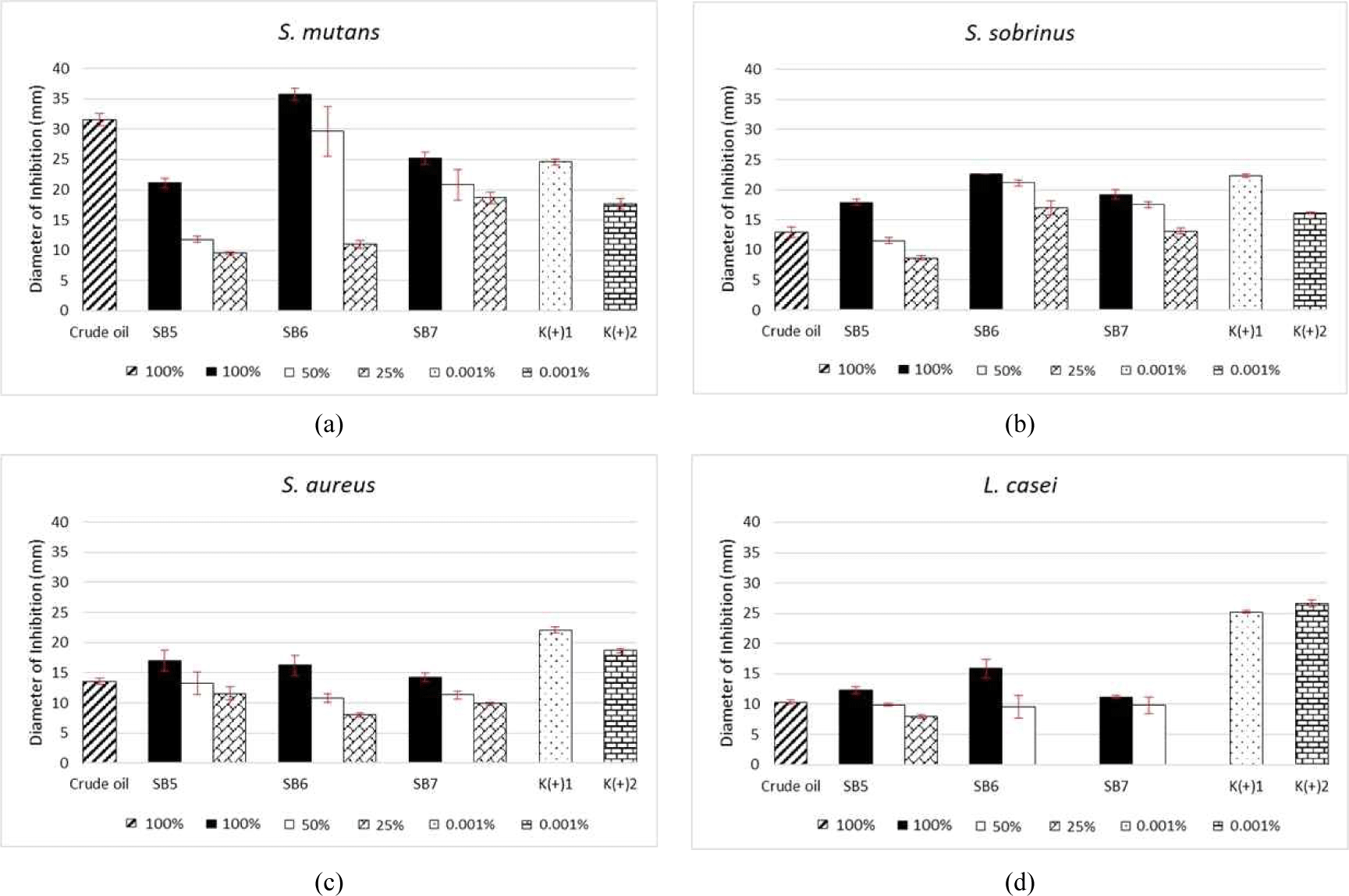
In analysis of the antibacterial activity against S. sobrinus, the largest diameter was observed for fraction SB6 (22.67 mm), followed by fractions SB7 (19.22 mm) and SB5 [17.89 mm; Fig. 2(a)]. Only the pure SB6 fraction surpassed the inhibition ability of both control antibiotics, whereas the other two pure fractions showed higher effects than chlorhexidine. All tested samples showed activity at a 50% concentration; the diameters from highest to lowest were SB6 > SB7 > SB5. These fractions, produced by SB distillation, inhibited S. sobrinus at up to 25%, with fraction SB5 achieving the smallest inhibition value (8.67 mm). Fraction SB6 was rich in chemical components of the monoterpene group, which strongly inhibits S. sobrinus. Alipour et al. (2015) reported that the strong antibacterial effect of essential oil from Ferula cupularis flowers, leaves, and stems was caused by the high content of hydrocarbon monoterpenes in such oils. Previous studies (El-Shenawy et al., 2015; Kotan et al., 2007; Zhu et al., 2013) showed that an essential oil with a high oxygenated monoterpene content was a strong antimicrobial agent.

As shown in Fig. 2(c), the lowest-concentration fractions of L. angulata (25%) generated using SB distillation actively inhibited S. aureus. The largest inhibition value was observed for fraction SB5 (17.00 mm); however, none of the fractions surpassed the effects of the positive controls. The second-largest value was observed for SB6, followed by SB7. The SB5 fraction contained some main compounds, namely α-phellandrene and p-cymene, from the monoterpene group, which are widely known for their pharmacological properties. Some monoterpenes, such as (+)-enantiomer, α-pinene, and β-pinene, exhibit antibacterial activity against C. albicans, Cryptococcus neoformans, Rhizopus oryzae, and methicillin-resistant S. aureus (da Silva et al., 2012). The essential oil from Litsea plant (i.e., L. cubeba) exhibits activity against S. aureus (Hu et al., 2019; Kačániová et al., 2020; Su and Ho, 2016; Thielmann et al., 2019; Van et al., 2016).

In examination of L. casei [Fig. 2(d)], the largest inhibition value was found in the SB6 fraction (15.89 mm), which remained active at 50% concentration. However, at a concentration of 25%, SB6 and SB7 showed no activity. The activity of the three fractions did not exceed that of the two control antibiotics. Essential oils, which contain members of the monoterpene group as the main compounds, exhibit antimicrobial activity against several strains of foodborne pathogens, both gram-positive and gram-negative, as well as against useful Lactobacilli strains (De Martino et al., 2009). In addition to containing α-phellandrene and p-cymene as the main compounds, fraction SB6 also contains ascaridole, which possesses anti-leishmanial (Monzote et al., 2018) and antimicrobial activities against phytopathogenic fungi and bacteria (Zefzoufi et al., 2020).

Thus, based on evaluation of the antibacterial activity from the pure fractions of L. angulata, fraction SF1, generated using the SF distillation method, showed the largest inhibition value, although the activities of fraction SB6 (collected from SB distillation) against S. mutans were insignificantly higher.

Based on evaluation of the antibacterial activity of the pure fractions of L. angulata, fraction SF1, prepared using SF distillation, had the largest inhibition value, although fraction SB6 (collected from SB distillation) showed slightly better activity against S. mutans. Fraction SF2 also had higher activity against L. casei, but the difference was less than 1 mm compared to that of SF1; thus, SF1 more effectively inhibited the four tested microorganisms, as it contained many hydrocarbon monoterpene compounds. The primary compound was α-phellandrene, which constituted over 40% of the sample. This compound exhibits various biological activities, including antibacterial, antifungal, and anticancer properties.

The antibacterial effects of different parts of L. angulata leaves towards the four tested bacteria more highly inhibited bacterial growth than the unseparated crude oil. Thus, separated essential oil is more effective than crude oil in inhibiting bacteria. Some studies indicated that separating an extract into fractions or single compounds leads to greater effects because the active compounds in plant extracts cannot synergize before separation by chromatography or other methods (Ahmad et al., 2011; Chua et al., 2019; Nguyen et al., 2017; Okwuchi, 2015; Onasanwo et al., 2008; Sannigrahi et al., 2010).

The size of the inhibition zone against microorganisms depends on the constituent chemical components in an essential oil, number of main components, concentration of the essential oil, and type and number of test bacteria (Baydar et al., 2004; Nazzaro et al., 2013). Essential oil disrupts the cell architecture first, causing damage to the membrane integrity and increasing permeability, which may interfere with numerous cellular activities. Essential oils can pass through the cell wall and cytoplasmic membrane and may disrupt or disturb different the fatty acid composition, phospholipid bilayer, and polysaccharide molecules. These changes may result in coagulation of the inner cell components of the cytoplasm and disrupt the bond between the fat and protein layer (Swamy et al., 2016). The release of cellular constituents in treated bacteria prompted the hypothesis that the primary effect of essential oils is membrane breakdown. Nonetheless, certain interactions with additional bacterial cell targets may strongly contribute to the observed antibacterial activities of the essential oil (Fadli et al., 2012). The structure of a compound is correlated with its microscopic characteristics, such as the molecular structure and macroscopic/empirical properties which include biological and molecular activities (Lee et al., 1996). Interactions between medicinal compounds and biological systems highly depend on the physical and chemical characteristics of the medicinal compounds, which are influenced by the types, amounts, and bonds between atoms, as well as the arrangement of the atomic space in the molecule (Utomo et al., 2017).

4. CONCLUSIONS

Essential oils from L. angulata leaves can be separated into fractions using both SF and SB distillation. The SF distillation method, particularly during the initial 60 min, yielded the highest oil content and produced fractions rich in bioactive compounds, especially α-phellandrene. GC-MS analysis confirmed that monoterpenes were the dominant constituents of the most active fractions.

Among all tested fractions, SF1 and SB6 exhibited the highest antibacterial activity, particularly against S. mutans, S. sobrinus, and S. aureus, whereas SF2 was the most effective against L. casei. Although antibacterial activity was observed at high concentrations (up to 100%), which were higher than the common minimum inhibitory concentrations (typically 0.01%–10%), the results reveal the application potential of these oils. Further research is needed to assess these activities at lower concentrations and to isolate specific bioactive compounds.

These findings highlight the potential of L. angulata essential oil, particularly its monoterpene-rich fractions, as a natural antibacterial agent for use in oral and dental products. Further in vivo investigations are needed to assess the safety, cytotoxicity, and efficacy of these fractions to support their application in pharmaceutical and personal care formulations.

CONFLICT of INTEREST

No potential conflict of interest relevant to this article was reported.

ACKNOWLEDGMENT

This project was funded by the Program Hibah Islamic Development Bank (IDB) 2018 (No. 448/UN17.45/DL/2018) and the Ministry of Research, Technology, and Higher Education of the Republic of Indonesia (Grant No. 221/UN17.41/KL/2019).

REFERENCES

1.

Agrawal, N., Choudhary, A.S., Sharma, M.C., Dobhal, M.P. 2011. Chemical constituents of plants from the genus Litsea. Chemistry & Biodiversity 8(2): 223-243.

2.

Agulló, L., Romero-Silva, M.J., Domenech, M., Seeger, M. 2017. p-Cymene promotes its catabolism through the p-Cymene and the p-cumate pathways, activates a stress response and reduces the biofilm formation in Burkholderia xenovorans LB400. PLOS ONE 12(1): 1-24.

3.

Agusta, A. 2000. Essential Oil of Indonesia Tropical Plant. ITB Press, Bandung, Indonesia.

4.

Ahmad, M.S., Nasshorudin, D., Mamat, A.S. 2014. Novel closed system extraction of essential oil: Impact on yield and physical characterization. In: Paris, France, 4th International Conference on Biotechnology and Environmental Management, pp. 42-46.

5.

Ahmad, N., Shinwari, Z.K., Hussain, J., Ahmad, S., Abbas, G., Zada, M., Ahmad, N., Iqbal, J. 2011. Biological evaluation of the crude extracts/fractions of Nepeta leavigata and Nepeta kurramensis. Journal of Pharmacy Research 4(10): 3472-3474.

6.

Akmal, R., Kuswanto, K., Fahrunnisa, S., Rahmi, R.A., Bayanil, N.E.P., Nurliani, A. 2016. The spermicide effect from methanol extract of Kalangkala seed (Litsea angulata) to spermatozoa mice (Mus musculus). Journal of Agromedicine and Medical Science 2(2): 18-23.

7.

Alipour, Z., Taheri, P., Samadi, N. 2015. Chemical composition and antibacterial activity of the essential oils from flower, leaf and stem of Ferula cupularis growing wild in Iran. Pharmaceutical Biology 53(4): 483-487.

8.

Amer, A., Mehlhorn, H. 2006. Repellency effect of forty-one essential oils against Aedes, Anopheles, and Culex mosquitoes. Parasitology Research 99(4): 478-490.

9.

Amrullah, R., Nurjanah, S., Widyasanti, A., Muhaemin, M. 2017. Study on effect of reflux ratios on patchouli oil properties produced by fractional distillation. Jurnal Teknotan 11(2): 77-88.

10.

Arfan, M., Amin, H., Karamać, M., Kosińska, A., Wiczkowski, W., Amarowicz, R. 2008. Antioxidant activity of phenolic fractions of Litsea monopetala [Persimon-leaved Litsea] bark extract. Polish Journal of Food and Nutrition Sciences 58(2): 229-233.

11.

Ariyani, D., Mustikasari, K. 2010. The phytochemistry screening of methanol extract from Kalangkala (Litsea angulata) seeds. Jurnal Sains dan Terapan Kimia 4: 131-136.

12.

Azizan, N., Mohd Said, S., Abidin, Z.Z., Jantan, I. 2017. Composition and antibacterial activity of the essential oils of Orthosiphon stamineus Benth and Ficus deltoidea Jack against pathogenic oral bacteria. Molecules 22(12): 1-18.

13.

Bach, S.M., Fortuna, M.A., Attarian, R., de Trimarco, J.T., Catalán, C.A.N., Av-Gay, Y., Bach, H. 2011. Antibacterial and cytotoxic activities of the sesquiterpene lactones cnicin and onopordopicrin. Natural Product Communications 6(2): 163-166.

14.

Badawy, M.E.I., Marei, G.I.K., Rabea, E.I., Taktak, N.E.M. 2019. Antimicrobial and antioxidant activities of hydrocarbon and oxygenated monoterpenes against some foodborne pathogens through in vitro and in silico studies. Pesticide Biochemistry and Physiology 158: 185-200.

15.

Balagopal, S., Arjunkumar, R. 2013. Chlorhexidine: The gold standard antiplaque agent. Journal of Pharmaceutical Sciences and Research 5(12): 270-274.

16.

Barrero, A.F., Quílez Del Moral, J.F., Lara, A., Herrador, M.M. 2005. Antimicrobial activity of sesquiterpenes from the essential oil of Juniperus thurifera wood. Planta Medica 71(1): 67-71.

17.

Bayala, B., Bassole, I.H., Scifo, R., Gnoula, C., Morel, L., Lobaccaro, J.M.A., Simpore, J. 2014. Anticancer activity of essential oils and their chemical components: A review. American Journal of Cancer Research 4(6): 591-607.

18.

Baydar, H., Saǧdiç, O., Özkan, G., Karadoǧan, T. 2004. Antibacterial activity and composition of essential oils from Origanum, Thymbra and Satureja species with commercial importance in Turkey. Food Control 15(3): 169-172.

19.

Buitrago, D., Velasco, J., Díaz, T., Morales, A. 2012. Chemical composition and antibacterial activity of the essential oil of Monticalia imbricatifolia Schultz (Asteraceae). Revista Latinoamericana de Química 40(1): 13-18.

20.

Chaiya, A., Saraya, S., Chuakul, W., Temsiririrkkul, R. 2013. Screening for dental caries: Preventive activities of medicinal plants against Streptococcus mutans. Mahidol University Journal of Pharmaceutical Sciences 40(1): 9-17.

21.

Chang, Y.T., Chu, F.H. 2011. Molecular cloning and characterization of monoterpene synthases from Litsea cubeba (Lour.) Persoon. Tree Genetics & Genomes 7: 835-844.

22.

Chatzopoulou, P.S., Katsiotis, S.T. 1995. Procedures influencing the yield and the quality of the essential oil from Juniperus communis L. berries. Pharmaceutica Acta Helvetiae 70(3): 247-253.

23.

Cheng, H.I., Lin, W.Y., Duh, C.Y., Lee, K.H., Tsai, I.L., Chen, I.S. 2001. New cytotoxic butanolides from Litsea acutivena. Journal of Natural Products 64(12): 1502-1505.

24.

Cheng, M.J., Wang, T.A., Lee, S.J., Chen, I.S. 2010. A new butanolide and a new secobutanolide from Litsea lii var. nunkao-tahangensis. Natural Product Research 24(7): 647-656.

25.

Chowdhury, J.U., Bhuiyan, M.N.I., Nandi, N.C. 2008. Aromatic plants of Bangladesh: Essential oils of leaves and fruits of Litsea glutinosa (Lour.) C.B. Robinson. Bangladesh Journal of Botany 37(1): 81-83.

26.

Chua, L.S., Lau, C.H., Chew, C.Y., Dawood, D.A.S. 2019. Solvent fractionation and acetone precipitation for crude saponins from Eurycoma longifolia extract. Molecules 24(7):1416.

27.

Crozier, A., Clifford, M.N., Ashihara, H. 2006. Plant Secondary Metabolites: Occurrence, Structure and Role in the Human Diet. Blackwell, Oxford, UK.

28.

Cruz, S.M., Marroquín, M.N., Gaitán, I.C., Cáceres, A. 2014. Antimicrobial activity of essential oils and ethanolic extracts of three species of laurel (Litsea spp.) from Guatemala. Acta Horticulturae 1030: 23-29.

29.

da Silva, A.C.R., Lopes, P.M., de Azevedo, M.M.B., Costa, D.C.M., Alviano, C.S., Alviano, D.S. 2012. Biological activities of a-pinene and β-pinene enantiomers. Molecules 17(6): 6305-6316.

30.

De Cássia da Silveira e Sá, R., Andrade, L.N., De Sousa, D.P. 2013. A review on anti-inflammatory activity of monoterpenes. Molecules 18(1): 1227-1254.

31.

De Martino, L., De Feo, V., Fratianni, F., Nazzaro, F. 2009. Chemistry, antioxidant, antibacterial and antifungal activities of volatile oils and their components. Natural Product Communications 4(12): 1741-1750.

32.

del Carmen Jiménez-Pérez, N., Lorea-Hernández, F.G., Jankowski, C.K., Reyes-Chilpa, R. 2011. Essential oils in Mexican Bays (Litsea spp., Lauraceae): Taxonomic assortment and ethnobotanical implications. Economic Botany 65: 178-189.

33.

Diastuti, H., Maolana Syah, Y., Dewi Juliawaty, L., Singgih, M. 2014. Antibacterial activity of germacrane type sesquiterpenes from Curcuma heyneana rhizomes. Indonesian Journal of Chemistry 14(1): 32-36.

34.

El-Shenawy, M.A., Baghdadi, H.H., El-Hosseiny, L.S. 2015. Antibacterial activity of plants essential oils against some epidemiologically relevant food-borne pathogens. The Open Public Health Journal 8: 30-34.

35.

Essien, E.E., Ogunwande, I.A., Setzer, W.N., Ekundayo, O. 2012. Chemical composition, antimicrobial, and cytotoxicity studies on S. erianthum and S. macranthum essential oils. Pharmaceutical Biology 50(4): 474-480.

36.

Fadli, M., Saad, A., Sayadi, S., Chevalier, J., Mezrioui, N.E., Pagès, J.M., Hassani, L. 2012. Antibacterial activity of Thymus maroccanus and Thymus broussonetii essential oils against nosocomial infection: Bacteria and their synergistic potential with antibiotics. Phytomedicine 19(5): 464-471.

37.

Feng, T., Zhang, R.T., Tan, Q.G., Zhang, X.Y., Liu, Y.P., Cai, X.H., Luo, X.D. 2009. Two new isoquinoline alkaloids from Litsea cubeba. Zeitschrift fur Naturforsch B 64(7): 871-874.

38.

Forssten, S.D., Björklund, M., Ouwehand, A.C. 2010. Streptococcus mutans, caries and simulation models. Nutrients 2(3): 290-298.

39.

Garcia, R., Alves, E.S.S., Santos, M.P., Viégas Aquije, G.M.F., Fernandes, A.A.R., dos Santos, R.B., Ventura, J.A., Fernandes, P.M.B. 2008. Antimicrobial activity and potential use of monoterpenes as tropical fruits preservatives. Brazilian Journal of Microbiology 39(1): 163-168.

40.

Geroushi, A., Auzi, A.A., Elhwuegi, A.S., Elzawam, F., Elsherif, A., Nahar, L., Sarker, S.D. 2011. Antiinflammatory sesquiterpenes from the root oil of Ferula hermonis. Phytotherapy Research 25(5): 774-777.

41.

Ghasemi Pirbalouti, A., Gholipour, Z. 2016. Chemical composition, antimicrobial and antioxidant activities of essential oil from Echinophora cinerea harvested at two phenological stages. Journal of Essential Oil Research 28(6): 501-511.

42.

Gogoi, R., Loying, R., Sarma, N., Munda, S., Kumar Pandey, S., Lal, M. 2018. A comparative study on antioxidant, anti-inflammatory, genotoxicity, anti- microbial activities and chemical composition of fruit and leaf essential oils of Litsea cubeba Pers from North-East India. Industrial Crops and Products 125: 131-139.

43.

Guzmán-Gutiérrez, S.L., Gómez-Cansino, R., García-Zebadúa, J.C., Jiménez-Pérez, N.C., Reyes-Chilpa, R. 2012. Antidepressant activity of Litsea glaucescens essential oil: Identification of β-pinene and linalool as active principles. Journal of Ethnopharmacology 143(2): 673-679.

44.

Hapsari, I.P., Feroniasanti, Y.M.L. 2019. Phytochemical screening and in vitro antibacterial activity of sweet basil leaves (Ocimum basilicum L.) essential oil against Cutibacterium acnes ATCC 11827. AIP Conference Proceedings 2099: 020007.

45.

Haque, T., Uddin, M.Z., Saha, M.L., Mazid, M.A., Hassan, M.A. 2014. Propagation, antibacterial activity and phytochemical profiles of Litsea glutinosa (Lour.) C. B. Robinson. Dhaka University Journal of Biological Sciences 23(2): 165-171.

46.

Ho, C.L., Hsu, K.P., Tseng, Y.H., Liao, P.C., Wang, E.I.C., Jeyashoke, N., Chien, T.C., Dong, W.C., Su, Y.C. 2010. Composition and antifungal activities of the leaf essential oil of Litsea coreana from Taiwan. Natural Product Communications 5(10): 1677-1680.

47.

Hosamath, P.V. 2011. Evaluation of antimicrobial activity of Litsea glutinosa. International Journal of Pharmaceutical Applications 2(1): 105-114.

48.

Hu, W., Li, C., Dai, J., Cui, H., Lin, L. 2019. Antibacterial activity and mechanism of Litsea cubeba essential oil against methicillin-resistant Staphylococcus aureus (MRSA). Industrial Crops and Products 130: 34-41.

49.

Indrawati, I., Anggraini, A.P., Irawan, B. 2017. Dental caries bacterial isolates sensitivity toward water-, ethanolic extract and essential oils of several shallot cultivars (Allium cepa). Nusantara Bioscience 9(2): 229-235.

50.

İşcan, G. 2017. Antibacterial and anticandidal activities of common essential oil constituents. Records of Natural Products 11(4): 374-388.

51.

Işcan, G., Kırımer, N., Demirci, F., Demirci, B., Noma, Y., Başer, K.H.C. 2012. Biotransformation of ()-(R)-α-phellandrene: Antimicrobial activity of its major metabolite. Chemistry & Biodiversity 9(8): 1525-1532.

52.

Jahangirian, H., Haron, M.J., Ismail, M.H.S., Rafiee-Moghaddam, R., Afsah-Hejri, L., Abdollahi, Y., Rezayi, M., Vafaei, N. 2013. Well diffusion method for evaluation of antibacterial activity. Digest Journal of Nanomaterials and Biostructures 8(3): 1263-1270.

53.

Jawetz, E., Melnick, J.L., Adelberg, E.A. 2005. Medical Microbiology. 1st ed. Salemba Medika, Jakarta, Indonesia.

54.

Jose, R.A., George Thengumpallil, S., Thushar, T.K.V. 2015. Litsea udayanii (Lauraceae): A new species from the southern western Ghats, India. Phytotaxa 222(1): 44-50.

55.

Kačániová, M., Terentjeva, M., Štefániková, J., Žiarovská, J., Savitskaya, T., Grinshpan, D., Kowalczewski, P.Ł., Vukovic, N., Tvrdá, E. 2020. Chemical composition and antimicrobial activity of selected essential oils against Staphylococcus spp. isolated from human semen. Antibiotics 9(11): 1-21.

56.

Kaluđerović, G.N., Krajnović, T., Momcilovic, M., Stosic-Grujicic, S., Mijatović, S., Maksimović-Ivanić, D., Hey-Hawkins, E. 2015. Ruthenium(II) p-cymene complex bearing 2,2′-dipyridylamine targets caspase 3 deficient MCF-7 breast cancer cells without disruption of antitumor immune response. Journal of Inorganic Biochemistry 153: 315-321.

57.

Kotan, R., Kordali, S., Cakir, A. 2007. Screening of antibacterial activities of twenty-one oxygenated monoterpenes. Zeitschrift fur Naturforsch C 62(7-8): 507-513.

58.

Kumar, R., Tripathi, Y.C. 2011. Getting Fragrance from Plants. Training Manual on Extraction Technology of Natural Dyes & Aroma Therapy and Cultivation Value Addition of Medicinal Plants. Forest Research Institute, Dehradun, India.

59.

Kuspradini, H., Putri, A.S., Diana, R. 2020. Toxicity, antioxidant ability and inhibition of oral pathogens by monoterpene-rich essential oil of Litsea angulata Blume. Agriculture and Natural Resources 54: 223-228.

60.

Kuspradini, H., Putri, A.S., Mitsunaga, T. 2018. Chemical composition, antibacterial and antioxidant activities of essential oils of Dryobalanops lanceolata Burck. Leaf. Research Journal of Medicinal Plant 12(1): 19-25.

61.

Kuspradini, H., Putri, A.S., Sukaton, E., Mitsunaga, T. 2016. Bioactivity of essential oils from leaves of Dryobalanops lanceolata, Cinnamomum burmannii, Cananga odorata, and Scorodocarpus borneensis. Agriculture and Agricultural Science Procedia 9: 411-418.

62.

Kuspradini, H., Wulandari, I., Putri, A.S., Tiya, S.Y., Kusuma, I.W. 2019. Phytochemical, antioxidant and antimicrobial properties of Litsea angulata extracts. F1000Research 7: 1839.

63.

Lee, K.W., Kwon, S.Y., Hwang, S., Lee, J.U., Kim, H. 1996. Quantitative structure-activity relationships (QSAR) study on C-7 substituted Quinolone. Bulletin of the Korean Chemical Society 17(2): 147-152.

64.

Lee, S.Y., Min, B.S., Kim, J.H., Lee, J., Kim, T.J., Kim, C.S., Kim, Y.H., Lee, H.K. 2005. Flavonoids from the leaves of Litsea japonica and their anti-complement activity. Phytotherapy Research 19(4): 273-276.

65.

Li, W.R., Shi, Q.S., Liang, Q., Xie, X.B., Huang, X.M., Chen, Y.B. 2014. Antibacterial activity and kinetics of Litsea cubeba oil on Escherichia coli. PLOS ONE 9(11): e110983.

66.

Li, X., Wong, C.H., Ng, T.W., Zhang, C.F., Leung, K.C.F., Jin, L. 2016. The spherical nanoparticle-encapsulated chlorhexidine enhances anti-biofilm efficiency through an effective releasing mode and close microbial interactions. International Journal of Nanomedicine 11: 2471-2480.

67.

Lin, J.J., Lu, K.W., Ma, Y.S., Tang, N.Y., Wu, P.P., Wu, C.C., Lu, H.F., Lin, J.G., Chung, J.G. 2014. Alpha-phellandrene, a natural active monoterpene, influences a murine WEHI-3 leukemia model in vivo by enhancing macrophague phagocytosis and natural killer cell activity. In Vivo 28(4): 583-588.

68.

Luo, M., Jiang, L.K., Huang, Y.X., Xiao, M., Li, B., Zou, G.L. 2004. Effects of citral on Aspergillus flavus spores by quasi-elastic light scattering and multiplex microanalysis techniques. Acta Biochimica et Biophysica Sinica 36(4): 277-283.

69.

Maiwald, L., Schwantes, P.A. 1991. Curcuma xanthorrhiza Roxb. Zeitschrift für Phytherapie 12: 35-45.

70.

Mangun, H.M.S., Waluyo, H., Purnama, A. 2005. Patchouli. Penebar Swadaya Grup, Depok, Indonesia.

71.

Marei, G.I., Rabea, E.I., Badawy, M.E.I. 2019. In vitro antimicrobial and antioxidant activities of monoterpenes against some food-borne pathogens. Journal of Plant Protection and Pathology 10(1): 87-94.

72.

Mathur, S., Mathur, T., Srivastava, R., Khatri, R. 2011. Chlorhexidine: The gold standard in chemical plaque control. National Journal of Physiology, Pharmacy & Pharmacology 1(2): 45-50.

73.

McCaig, L.F., McDonald, L.C., Mandal, S., Jernigan, D.B. 2006. Staphylococcus aureus–associated skin and soft tissue infections in ambulatory care. Emerging Infectious Diseases 12(11): 1715-1723.

74.

Monzote, L., Geroldinger, G., Tonner, M., Scull, R., De Sarkar, S., Bergmann, S., Bacher, M., Staniek, K., Chatterjee, M., Rosenau, T., Gille, L. 2018. Interaction of ascaridole, carvacrol, and caryophyllene oxide from essential oil of Chenopodium ambrosioides L. with mitochondria in Leishmania and other eukaryotes. Phytotherapy Research 32(9): 1729-1740.

75.

Nazzaro, F., Fratianni, F., De Martino, L., Coppola, R., De Feo, V. 2013. Effect of essential oils on pathogenic bacteria. Pharmaceuticals 6(12): 1451-1474.

76.

Nguyen, V.T., Sakoff, J.A., Scarlett, C.J. 2017. Physicochemical properties, antioxidant and cytotoxic activities of crude extracts and fractions from Phyllanthus amarus. Medicines 4(2): 42.

77.

Niksic, H., Kovac-Besovic, E., Omeragic, E., Muratovic, S., Kusturica, J., Duric, K. 2017. Antiproliferative, antimicrobial, and antioxidant activity of Lavandula angustifolia Mill. essential oil. Journal of Health Sciences 7(1): 35-43.

78.

Nurainy, F., Rizal, S., Yudiantoro. 2008. The effect of chitosan concentrations on the antibacterial activity with gel diffusion/well method. Jurnal Teknologi & Industri Hasil Pertanian 13(2): 117-125.

79.

Okwuchi, N.P. 2015. Extraction, fractionation and assessment of antioxidant activities of active components of Aframomum sceptrum seeds. African Journal of Biochemistry Research 9(10): 117-123.

80.

Oliveira, J., Alves, D.K.M., Miranda, M.L.D., Alves, J.M., Xavier, M.N., Cazal, C.M., Alves, C.C.F. 2017. Chemical composition of essential oil extracted from leaves of Campomanesia adamantium subjected to different hydrodistillation times. Ciência Rural 47(1): 1-7.

81.

Onasanwo, S.A., Pal, A., George, B., Olaleye, S.B. 2008. Pharmacological and toxicity studies of the crude extracts and fractions of Hedranthera barteri leaf in rats and mice. African Journal of Biomedical Research 11(3): 311-321.

82.

Pambudi, W.A., Sukaton, E., Kuspradini, H. 2018. Antifungal activity of Litsea rubiginosa n-hexane extract. In: Samarinda, Indonesia, Proceedings of the National Seminar, Faculty of Agricultural Products Technology, Mulawarman University, pp. 158-164.

83.

Pan, J.Y., Zhang, S., Wu, J., Li, Q.X., Xiao, Z.H. 2010. Litsea glutinan A and lignans from Litsea glutinosa. Helvetica Chimica Acta 93(5): 951-957.

84.

Păunescu, E., Nowak-Sliwinska, P., Clavel, C.M., Scopelliti, R., Griffioen, A.W., Dyson, P.J. 2015. Anticancer organometallic osmium(II)-p-cymene complexes. ChemMedChem 10(9): 1539-1547.

85.

Prins, C.L., Lemos, C.S.L., Freitas, S.P. 2006. Efeito do tempo de extração sobre a composição e o rendimento do óleo essencial de alecrim (Rosmarinus officinalis). Revista Brasileira de Plantas Medicinais 8(4): 92-95.

86.

Putri, A.S., Purba, F.F., Kusuma, I.W., Kuspradini, H. 2018. Chemical compositions and antimicrobial potential of Actinodaphne macrophylla leaves oils from East Kalimantan. IOP Conference Series: Earth and Environmental Science 144:012021.

87.

Qin, J., Wang, J., Yao, Y. 2014. Study on polymerization inhibitor of isoprene during extractive distillation process. Speciality Petrochemicals 31(6).

88.

Rahimmalek, M., Goli, S.A.H. 2013. Evaluation of six drying treatments with respect to essential oil yield, composition and color characteristics of Thymys daenensis subsp. daenensis. Celak leaves. Industrial Crops and Products 42: 613-619.

89.

Rath, B. 2005. Medicina contributions from some species of Litsea. The Indian Forester 131: 479-482.

90.

Saddiq, A.A., Khayyat, S.A. 2010. Chemical and antimicrobial studies of monoterpene: Citral. Pesticide Biochemistry and Physiology 98(1): 89-93.

91.

Sannigrahi, S., Mazuder, U.K., Pal, D.K., Parida, S., Jain, S. 2010. Antioxidant potential of crude extract and different fractions of Enhydra fluctuans Lour. Iranian Journal of Pharmaceutical Research 9(1): 75-82.

92.

Saputri, R., Susiani, E.F. 2018. Antioxidant activity test of ethanolic extract of Kalangkala (Litsea angulata) fruits and seeds from South Kalimantan. Borneo Journal of Pharmacy 1(2): 81-84.

93.

Savakar, P.P., Kharade, S.G., Talap, P.P., Moharir, S.R., Suryawanshi, M.A. 2017. Study of spinning band distillation column. International Journal of Advance Engineering and Research Development 4(4): 1060-1065.

94.

Silva, V.A., Sousa, J.P., Guerra, F.Q.S., Pessôa, H.L.F., Freitas, A.F.R., Coutinho, H.D.M., Alves, L.B.N., Lima, E.O. 2015. Antibacterial activity of the monoterpene linalool: Alone and in association with antibiotics against bacteria of clinical importance. International Journal of Pharmacognosy and Phytochemical Research 7: 1022-1026.

95.

Sinaga, A. 2013. Factors related to maternal behavior in preventing tooth decay in children aged 1-5 years at Babakan Sari Health Center Bandung. Jurnal Darma Agung XXI: 1-10.

96.

Sousa, R.M.F., de Morais, S.A.L., Vieira, R.B.K., Napolitano, D.R., Guzman, V.B., Moraes, T.S., Cunha, L.C.S., Martins, C.H.G., Chang, R., de Aquino, F.J.T., do Nascimento, E.A., de Oliveira, A. 2015. Chemical composition, cytotoxic, and antibacterial activity of the essential oil from Eugenia calycina Cambess. leaves against oral bacteria. Industrial Crops and Products 65: 71-78.

97.

Stratakos, A.C., Koidis, A. 2015. Methods for Extracting Essential Oils. In: Essential Oils in Food Preservation, Flavor and Safety, Ed. by Preedy, V.R. CRC Press, Boca Raton, FL, USA.

98.

Su, P.W., Yang, C.H., Yang, J.F., Su, P.Y., Chuang, L.Y. 2015. Antibacterial activities and antibacterial mechanism of Polygonum cuspidatum extracts against nosocomial drug-resistant pathogens. Molecules 20(6): 11119-11130.

99.

Su, Y.C., Ho, C.L. 2016. Essential oil compositions and antimicrobial activities of various parts of Litsea cubeba from Taiwan. Natural Product Communications 11(4): 515-518.

100.

Supriono, Susanti, T.A. 2014. The quality of Patchouli essential oil from the size reduction method in patchouli plant distillation (Pogostemom cablin BENTH). In: Jatinangor, Indonesia, Prosiding Seminar Nasional Kimia, pp. 1-6.

101.

Surono, I. 2004. Probiotics, Fermented Milk and Health. PT. Tri Cipta Karya, Jakarta, Indonesia.

102.

Swamy, M.K., Akhtar, M.S., Sinniah, U.R. 2016. Antimicrobial properties of plant essential oils against human pathogens and their mode of action: An updated review. Evidence-based Complementary and Alternative Medicine 2016: 3012462.

103.

Tanaka, H., Yatsuhashi, S., Yasuda, T., Sato, M., Sakai, E., Xiao, C., Murata, H., Murata, J. 2009. A new amide from the leaves and twigs of Litsea auriculata. Journal of Natural Medicines 63(3): 331-334.

104.

Thielmann, J., Muranyi, P., Kazman, P. 2019. Screening essential oils for their antimicrobial activities against the foodborne pathogenic bacteria Escherichia coli and Staphylococcus aureus. Heliyon 5(6): e01860.

105.

Thongthip, P., Atiphasaworn, P., Monggoot, S., Pansanit, A., Pripdeevech, P. 2017. In vitro antibacterial activity and essential oil composition of Litsea petiolata Hook. f. leaves. Journal of Applied Pharmaceutical Science 7(09): 094-098.

106.

Tisserand, R., Young, R. 2013. Essential Oil Safety: A Guide for Health Care Professionals. Elsevier Health Sciences, London, UK.

107.

Torres, F.C., Lucas, A.M., Ribeiro, V.L.S., Martins, J.R., von Poser, G., Guala, M.S., Elder, H.V., Cassel, E. 2012. Influence of essential oil fractionation by vacuum distillation on acaricidal activity against the cattle tick. Brazilian Archives of Biology and Technology 55(4): 613-621.

108.

Turek, C., Stintzing, F.C. 2013. Stability of essential oils: A review. In Comprehensive Reviews in Food Science and Food Safety 12(1).

109.

U.S. Department of Health and Human Services. 2000. Final Report on Carcinogens Background Document for Chloramphenicol. Meeting of the NTP Board of Scientific Counselors Report on Carcinogens Subcommittee. U.S. Department of Health and Human Services, Washington, DC, USA.

110.

Utomo, S.B., Sanubari, F., Utami, B., Nurhayati, N.D. 2017. Analysis of a quantitative relationship between the structure and analgesic activity of meperidin derivatives using semi-empirical AM1 method. Jurnal Kimia dan Pendidikan Kimia 2(3): 158-168.

111.

van Den Dool, H., Dec. Kratz, P. 1963. A generalization of the retention index system including linear temperature programmed gas: Liquid partition chromatography. Journal of Chromatography A 11: 463-471.

112.

Van, N.H., Nguyen, V.T.H., Trang, V.T., Tien, P.Q., Nhan, K.T., Sarter, S., Son, C.K. 2016. Antimicrobial activities and interaction effects of Vietnamese Litsea cubeba (Lour.) Pers essential oil and its endophytic actinobacteria. Journal of Science and Technology 54(4A): 234-241.

113.

Van Winkelhoff, A.J., Schouten-van Meeteren, A.Y.N., Baart, J.A., Vandenbroucke-Grauls, C.M.J.E. 2000. Microbiology of destructive periodontal disease in adolescent patients with congenital neutropenia: A report of 3 cases. Journal of Clinical Periodontology 27(11): 793-798.

114.

Wang, H., Liu, Y. 2010. Chemical composition and antibacterial activity of essential oils from different parts of Litsea cubeba. Chemistry & Biodiversity 7(1): 229-235.

115.

Wang, Y.S., Huang, R., Lu, H., Li, F.Y., Yang, J.H. 2010. A new 2′-oxygenated flavone glycoside from Litsea glutinosa (Lour.) C. B. Rob. Bioscience, Biotechnology, and Biochemistry 74(3): 652-654.

116.

Widayati, N. 2014. Factors associated with dental caries in children aged 4-6 years old. Jurnal Berkala Epidemiologi 2(2): 196-205.

117.

Wong, M.H., Lim, L.F., Ahmad, F., Assim, Z. 2014. Antioxidant and antimicrobial properties of Litsea elliptica Blume and Litsea resinosa Blume (Lauraceae). Asian Pacific Journal of Tropical Biomedicine 4(5): 386-392.

118.

Wulandari, I., Kusuma, I.W., Kuspradini, H. 2018. Antioxidant and antibacterial activity of Litsea garciae. IOP Conference Series: Earth and Environmental Science 144: 012024.

119.

Yang, T.S., Liou, M.L., Hu, T.F., Peng, C.W., Liu, T.T. 2013. Antimicrobial activity of the essential oil of Litsea cubeba on cariogenic bacteria. Journal of Essential Oil Research 25(2): 120-128.

120.

Yusuf, M., Begum, J., Hoque, M.N., Choudhury, J.U. 2009. Medicinal Plants of Bangladesh-revised and Enlarged. Bangladesh Council of Scientific and Industrial Research Laboratory, Chittagong, Bangladesh.

121.

Zefzoufi, M., Smaili, A., Fdil, R., Rifai, L.A., Faize, L., Koussa, T., Makroum, K., Ben Ali, A., Tabyaoui, M., Mouzdahir, A., Sraidi, K., Faize, M. 2020. Composition of essential oil of Moroccan Dysphania ambrosioides and its antimicrobial activity against bacterial and fungal phytopathogens. Journal of Plant Pathology 102(1): 47-58.

122.

Zhang, H.J., Tan, G.T., Santarsiero, B.D., Mesecar, A.D., Van Hung, N., Cuong, N.M., Soejarto, D.D., Pezzuto, J.M., Fong, H.H.S. 2003. New sesquiterpenes from Litsea verticillata. Journal of Natural Products 66(5): 609-615.

123.

Zhang, J., Sun, H., Chen, S., Zeng, L., Wang, T. 2017. Anti-fungal activity, mechanism studies on α-phellandrene and nonanal against Penicillium cyclopium. Botanical Studies 58(1): 13.

124.

Zhao, Y., Shong, G.Q., Guo, Y.W. 2003. Rotundifolinol, a new 1,3-diarylpropan-2-ol from bark of Litsea rotundifolia var. oblongifolia. Journal of Asian Natural Products Research 5(4): 273-277.

125.

Zheljazkov, V.D., Astatkie, T., Schlegel, V. 2014. Hydrodistillation extraction time effect on essential oil yield, composition, and bioactivity of coriander oil. Journal of Oleo Science 63(9): 857-865.

126.

Zhu, J., Lower-Nedza, A.D., Hong, M., Jiec, S., Wang, Z., Yingmao, D., Tschiggerl, C., Bucar, F., Brantner, A.H. 2013. Chemical composition and antimicrobial activity of three essential oils from Curcuma wenyujin. Natural Product Communications 8(4): 523-526.