
1. INTRODUCTION
Studies on wooden cultural properties begin with accurate wood identification. The reason is that wood itself is not only a record of various climate and environmental changes, but can also provide information to predict vegetation, social, cultural and trade relations of the past. General wood identification is performed by visual inspection of various wood tissues based on established wood anatomy (Eom and Park, 2018, Park et al., 2018; Lee et al., 2018). Although visual inspection is the most reliable method, there are practical limitations when sampling is extremely limited, such as for important wooden cultural properties.
Computed tomography (CT) is a non-destructive imaging technique that visualizes the internal threedimensional structure of wood by projecting X-rays to an object at various angles and reconstructing the absorption difference through mathematical methods using a computer. However, conventional CT imaging has failed to provide images by which anatomical characteristics can be observed from wood owing to its low spatial resolution. Computer vision-based methods should be used to identify wood species using CT images (Kobayashi et al., 2015, 2019a). Synchrotron X-ray microtomography (SR-μCT) combines the synchrotron X-ray micro-imaging with CT techniques and provides a spatial resolution of tens of nanometers to micrometers to visualize the structure inside a tiny object. SR-μCT has been considered as an alternative to the conventional method owing to the limiting of the wood sample size (Steppe et al., 2004; Van den Bulcke et al., 2009; Mizuno et al., 2010; Tazuru and Sugiyama, 2019).
This study presents three-dimensional microstructural images of small samples taken from Manseru pavilion in Bongjeongsa temple by SR-μCT and reports the identification results.
2. EXPERIMENTAL
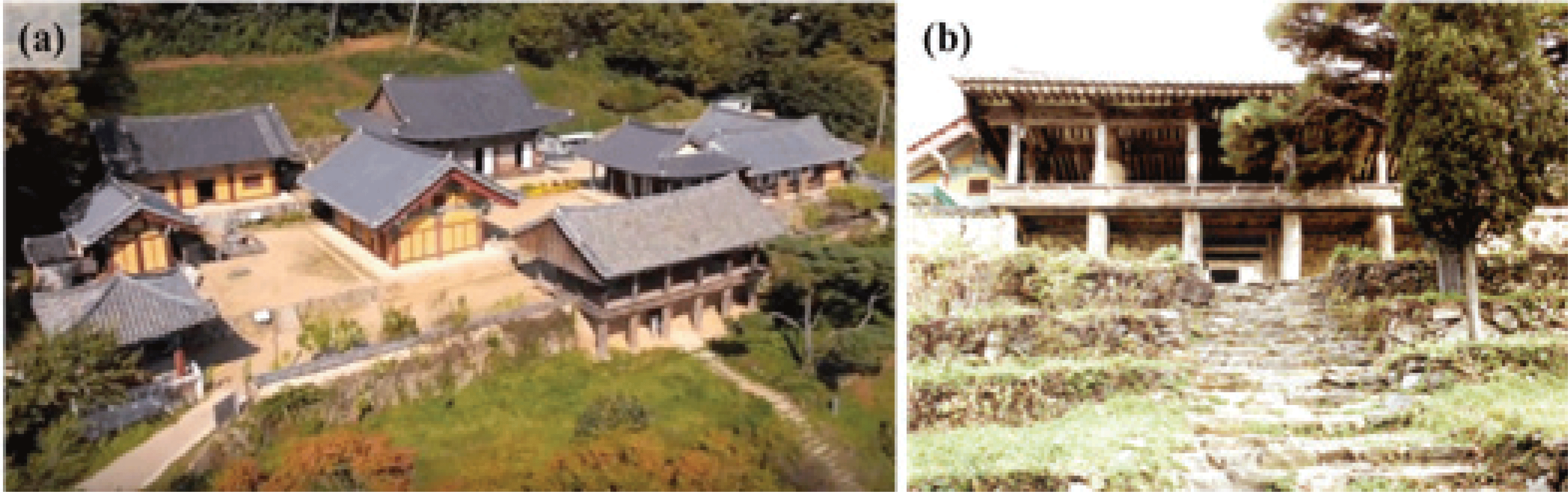
Four wood fragments collected from wooden members used in Manseru pavilion of Bongjeongsa temple, Andong, Korea, were provided from the Korea Forestry Promotion Institute in 2017. Manseru, the pavilion and main gate of Bongjeongsa temple, was built in 1680 and is designated as a tangible cultural property of Gyeongsangbuk-do Province. Moreover, Bongjeongsa temple is one of the seven South Korean Buddhist temples listed as a UNESCO World Heritage Site in 2018 under the name Sansa, Buddhist mountain monasteries in Korea. The samples were taken from the first two columns on the left side of the ground floor and from a beam and a joist on the second floor. The samples were tiny with a thin and long shape, ranging in size from a minimum of 2.2 × 1.7 × 7.2 mm (R × T × L) to a maximum of 1.4 × 5.8 × 13.4 mm.
To obtain three-dimensional microstructural images from the samples, the SR-μCT scan was performed on the beamline 20XU at SPring-8, Hyogo Prefecture, Japan. The synchrotron is a large ring-shaped electron accelerator, and electrons can achieve energies of several GeV. When electrons are forced to move out of their orbit by deflecting or bending magnets, they emit a synchrotron X-ray (Casali, 2006; Creagh, 2007). Three-dimensional imaging can be performed through computed tomography using the synchrotron X-ray. The beamline 20XU is designed to apply various imaging technologies such as microtomography, X-ray microscopy, and medical imaging. For a detailed technical description of this beamline, see the article by Suzuki et al. (2004).
Wood samples were cut and trimmed into a cylindrical shape with 5-mm height and 0.7-mm diameter and then fixed to a specially designed support stick for scanning. Two specimens from each sample, a total of eight specimens, were prepared. The mounted specimen was scanned with a stepwise rotation of 0.1 degree, and a total of 1800 projections were acquired by a CCD camera coupled with an optical lens. The size and resolution of the acquired images were 2048 × 2048 pixels and 0.472 μm/pixel, respectively. Then, 2048 slide images were reconstituted by an in-house developed software program ‘CT’.
The images were visualized and analyzed using the software VGStudio MAX 2.2 (Volume Graphics GmbH, Germany). The 2048 two-dimensional images for each sample were reconstructed to generate a three-dimensional image. The software allows for free cutting of the images, that is, it can observe all sections required by the user. Wood identification was carried out on the basis of the anatomical characteristics obtained by observing three orthogonal sections. All the reconstructed images were preprocessed with a median filter to remove noise.
3. RESULTS and DISCUSSION
Three-dimensional microstructural images of the four wood samples were obtained by SR-μCT with image reconstruction software. The images were clear enough to distinctly observe the anatomical characteristics present in the reconstructed area.
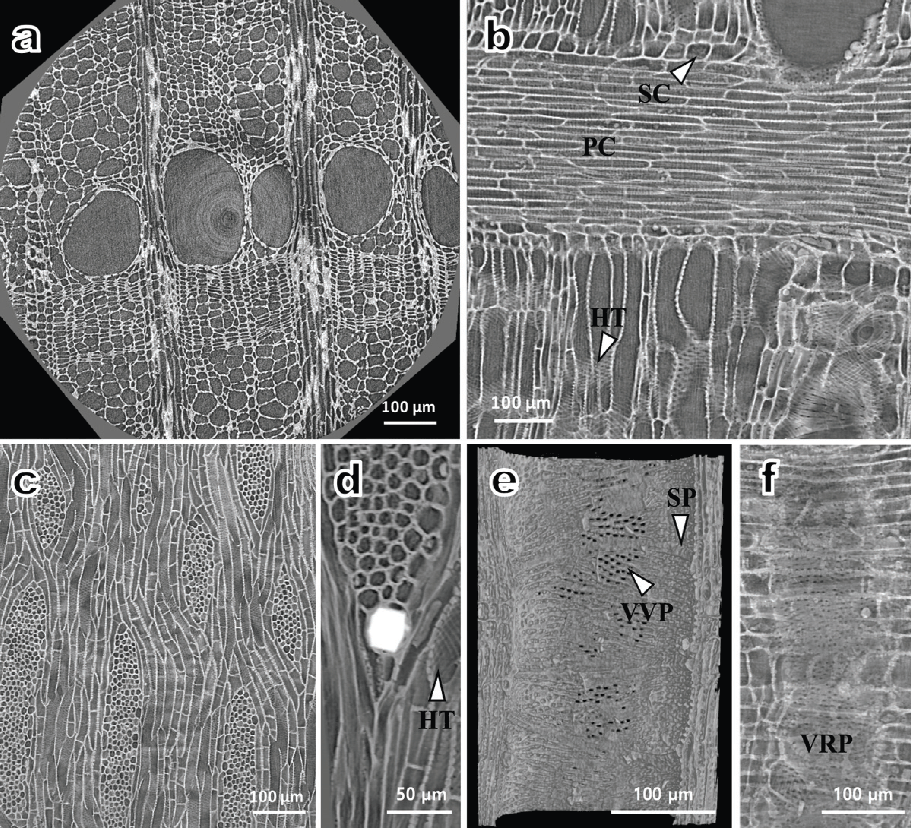
Fig. 2 presents reconstructed images of the wood samples taken from the first column. The growth ring boundary was distinct and the vessel arrangement was ring-porous with diameters ranging from 85 to 165 micrometers (Fig. 2a). One thing to note here is that the concentric shapes observed in the centers of Figs. 2a and 3a are ring artifacts commonly encountered in computed tomography and are not derived from wood. Ring artifacts are caused by a variety of factors, including miscalibration, detector failure, or insufficient radiation dose in a CT scanner (Triche et al., 2019). In the radial section (Fig. 2b), the cellular composition of rays consisted of procumbent cells with a row of square marginal cells. In addition, helical thickenings were only present in narrower vessel elements. Multiseriate rays with 2–9 seriate were observed in the tangential section (Fig. 2c), and crystals existed in enlarged cells (Fig. 2d). The arrangement of intervessel pits and vessel-ray parenchyma pits were both alternate type (Fig. 2e and 2f). Simple perforation plates were observed in the images obtained by cutting the vessels parallel to the axial direction. All anatomical evidence observed from the images was consistent with the characteristics of Zelkova serrata.
Reconstructed images of the second column are shown in Fig. 3. The porosity of vessels seemed to be diffuse-porous with a radial pattern in the cross section (Fig. 3a). Crystals were observed in some axial parenchyma cells. There were vasicentric tracheids around vessels in the radial section (Fig. 3b). In the tangential section (Fig. 3c), rays with two distinct sizes were observed. The uniseriate ray and the broad ray both contained crystals, and the cellular composition of rays were all procumbent cells. A tylosis and simple perforation plates were present in vessels (Fig. 3d). Furthermore, vessel-ray parenchyma pits were a vertically arranged palisade type (Fig. 3e). This is a genus specific feature of Quercus.
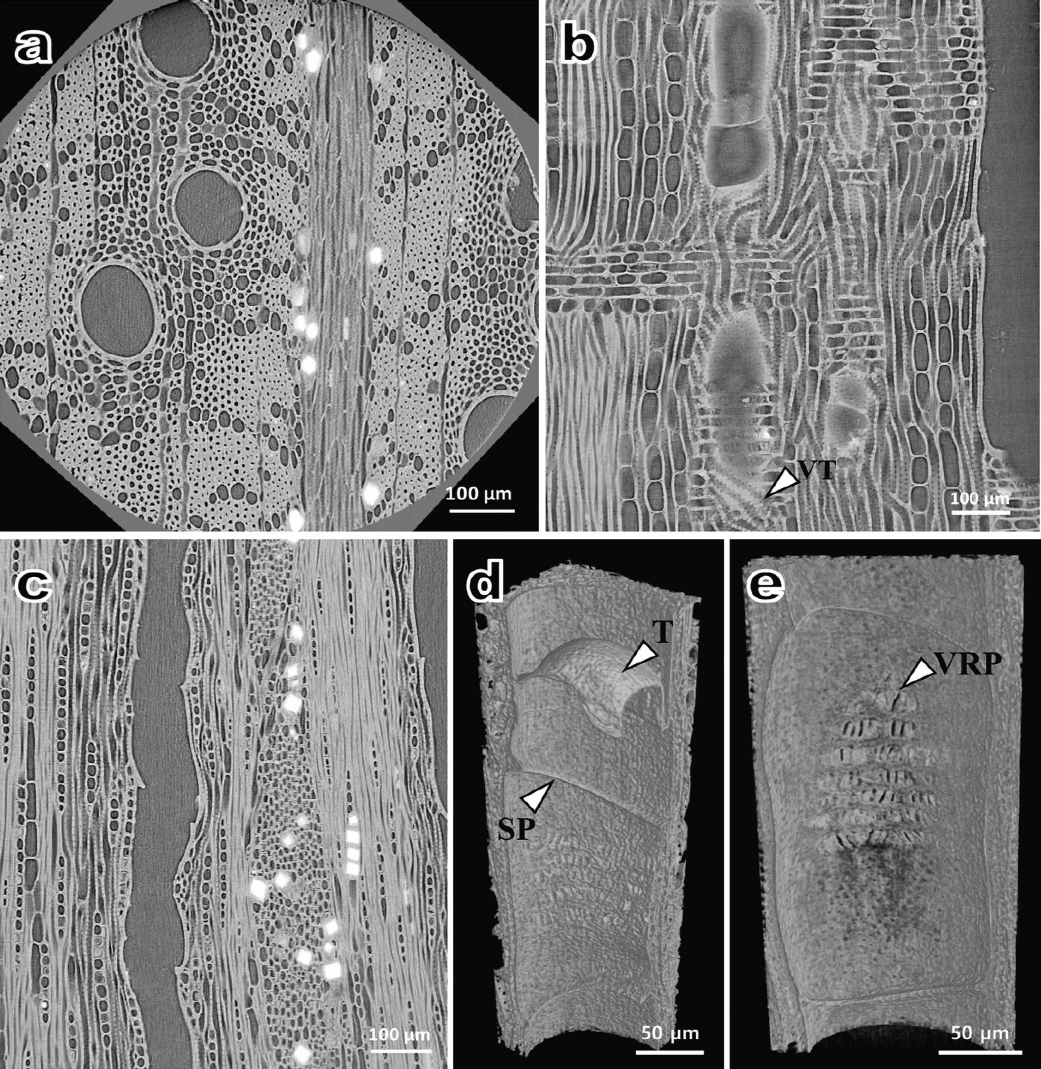
All the features observed in Fig. 3 suggest Quercus subg. Cyclobalanopsis (Noshiro and Sasaki, 2011; Kobayashi et al., 2019b). However, it is difficult to conclude that the porosity is diffuse porous with a radial pattern because the vessel arrangement in earlywood could not be observed in the cross section (Fig. 3a). Without considering wood porosity, all the features are also consistent with Quercus sect. Cerris. To determine the porosity of the sample, macroscopic visual inspection using a stereomicroscope was performed on other specimens taken from the same column. As a result, the sample was identified as a ring porous species owing to the presence of large earlywood vessels with tangential diameters greater than 200 μm in a specimen. From these observations, we classified the sample as Quercus sect. Cerris.
Fig. 4 presents the cross sections and rays of wood samples taken from the beam (Fig. 4a and 4b) and joist (Fig. 4c and 4d). In the cross section of the beam (Fig. 4a), an axial resin canal with thin-walled epithelial cells and a gradual transition from earlywood to latewood were observed. Cross-field pits were windowlike type and ray tracheids were present in rays (Fig. 4b). These features can be observed in Pinus spp. among the native species in Korea.

P. densiflora and P. thunbergii in the subgenus Diploxylon, and P. koraiensis, P. pumila, and P. parviflora in the subgenus Haploxylon, are representative Pinus species in Korea. The key criterion for classification between the Diploxylon and Haploxylon species is the presence or absence of dentate thickening in the ray tracheid wall. The Diploxylon species have dentate thickenings in ray tracheids, but these are not observed in Haploxylon. In Fig. 4b, the ray tracheids had smooth walls without dentate. Therefore, it was considered that the samples are close to the Haploxylon species, and the gradual transition from earlywood to latewood is another piece of evidence for Haploxylon (Fig. 4a).
Among the Haploxylon species, P. pumila, which belongs to the subsection Cembrae together with P. koraiensis, is not used as a structural member owing its small size. The smooth end wall of the ray parenchyma cell observed in Fig. 4b is an important key for distinguishing P. koraiensis from P. parviflora with distinctly pitted end walls (Eom, 2015). In addition, P. parviflora, a subsection Strobi, is a species only native to Ulleungdo Island, which is more than 200 km away, in a straight line, from Bongjeongsa temple, so it is very unlikely that the species was used in the construction of Manseru pavilion. For these reasons, the sample taken from the beam was judged to be P. koraiensis.
For the joist, there were no axial resin canals and axial parenchyma cells in the cross section (Fig. 4c). However, owing to the presence of window-like cross field pits and ray tracheids with a smooth wall in the radial section, this sample was also judged to be P. koraiensis.
From the wood identification of Manseru pavilion, two columns were identified as Zelkova serrata and Quercus spp., while both beam and joist were identified as Pinus koraiensis. At the time of the construction of Manseru pavilion (1680), the wood species identified in this study were commonly used to build Buddhist temples (Park and Lee, 2007). One interesting point here is that Pinus species in Diploxylon was used for the columns of Geungnakjeon and Daeungjeon Halls, the other major buildings in Bongjeongsa temple (Park et al., 2005). Considering the fact that the logging of the Pinus trees was strictly controlled in the Joseon Dynasty, the difference in wood species may reflect a social aspect at the time they were built. During the Joseon Dynasty (1392–1897), when the Manseru pavilion was built, Buddhism was oppressed as Confucianism was a national ideology. In contrast, Buddhism was a national religion during the Goryeo Dynasty (918–1392), when the Geungnakjeon and Daeungjeon Halls were built, which are estimated to be the thirteenth and fourteenth centuries, respectively.
4. CONCLUSION
Using SR-μCT, internal microstructures of wood samples that cannot be visualized by optical methods were reconstructed into three-dimensional images. The spatial resolution of this technique was sufficient to observe the major anatomical features of wood. The wood samples collected from the Manseru pavilion were identified as Zelkova serrata, Quercus sect. Cerris and Pinus koraiensis. The images reconstructed by SR-μCT are useful for obtaining various anatomical information owing to free manipulation such as cropping in various directions, extracting and modifying specific areas. There are many important wooden cultural properties that have not yet been identified owing to their managing specificity, especially for non-building wooden artifacts. SR-μCT is an effective method of identifying such wooden artifacts with minimal damage. Furthermore, if there are naturally delaminated small wood fragments that commonly occur during the preservation process or storage of the artifacts, SR-μCT will not involve any further damage for wood identification.